

Invasive urothelial carcinoma makes up only 1% to 2% of all neoplastic disorders in dogs. Patients present with nonspecific clinical signs of pollakiuria, stranguria, and hematuria and are commonly treated for urinary tract infections without further investigation. A cancer diagnosis can be scary to a client, and it is the job of veterinary professionals to provide support and care for the patient. Quality of life is the key goal in oncology in veterinary medicine. Sometimes that care includes not just treating the patient’s cancer but also treating the pain that may be associated with the neoplastic process. This case report reviews the treatment plan for a dog with advanced-stage invasive urothelial carcinoma at diagnosis.
Take-Home Points
- Invasive urothelial cell carcinoma makes up only 1% to 2% of all canine neoplastic disorders.
- Pollakiuria and stranguria do not always mean a mass is present in the urinary tract.
- At diagnosis of invasive urothelial carcinoma, 20% of patients will have distant metastasis.
- Quality of life is very important for the cancer patient and the owner.
- Patients with metastatic bone lesions can have a good quality of life when multimodal pain management is prescribed.
Invasive urothelial carcinoma (iUC), formerly known as transitional cell carcinoma of the urinary bladder, is the most common neoplasia of the urinary tract. It makes up 1% to 2% of all neoplastic disorders in the dog.1 Patients present with nonspecific clinical signs, such as dysuria, pollakiuria, and hematuria.
The initial test recommended for a patient with nonspecific urinary signs is a urinalysis. On the urinalysis, the presence of white blood cells and bacteria, specifically intracellular rods or cocci on the sediment, point to a urinary tract infection (UTI). When bacteria are noted on a urinalysis, a urine culture is recommended to test for the specific bacteria present and to assess what antibiotic the bacteria are susceptible to. If bacteria are not present on the urinalysis, other things to assess for are the presence of atypical epithelial cells, red blood cells, urinary crystals, and/or stones. Further workup is recommended when patients do not respond to antibiotic therapy or if there is no sign of a UTI on the urinalysis.
The most common location for iUC is the trigone of the urinary bladder. It may also extend into or affect the urethra. At the time of diagnosis, approximately 20% of patients present with metastatic disease, and at the time of death, 50% of patients will have distant metastasis.2
Primary Care
Lily was an 8-year-old female spayed Shih Tzu (FIGURE 1) that was initially presented to the referring veterinarian in July 2020 for pollakiuria and tenderness in the abdomen when the owner picked her up. The referring veterinarian performed frequent urinalyses that showed the presence of bacteria (both rods and cocci). The veterinarian recommended urine cultures each time, but they were not pursued for further investigation. Lily was treated off and on for 3 months for UTIs, with little to no response to antibiotic therapy. The veterinarian performed abdominal radiography when Lily continued to exhibit signs of UTI, and cystoliths were observed. The presence of cystoliths on radiographs fit with her clinical signs because cystoliths can cause irritation of the urinary bladder wall and discomfort. Cystoliths may also cause pollakiuria because their presence creates the sensation that the urinary bladder is not completely empty and/or they may be in an area of the urinary bladder that blocks the normal flow of urine. Due to the presence of cystoliths, a therapeutic urinary diet was recommended.
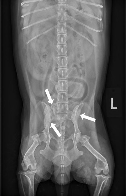
Lily’s clinical signs continued to worsen after initiation of the urinary diet. Repeat abdominal radiography was performed, along with abdominal ultrasonography. The radiographs revealed a periosteal reaction on the right wing of the ilium, a mass-like structure in the lumen of the urinary bladder, a thickened urethra, and enlargement of the medial iliac lymph nodes.

Figure 1. The patient, Lily. Courtesy Christopher Garber.
Referral Care
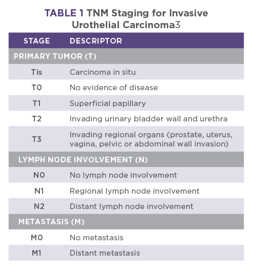
In October 2020, Lily was presented to the referral oncology service for workup of a urinary bladder mass with probable metastatic disease to the right ilium. Differential diagnosis included iUC with metastasis to the bone and iliac lymph nodes; neoplastic processes; and, less likely, an infectious process. The periosteal reaction of the bone may have been a primary bone tumor (e.g., osteosarcoma) but was more likely related to spread of the iUC. Osteosarcoma predominantly affects large-breed dogs and is most often found in the appendicular skeleton. The wing of the ilium is not a common site, and with the close location to the urinary bladder, the concern was that Lily’s cancer had spread to her bone. Her TNM (tumor, node, and metastasis) stage was diagnosed as T3N1M1 (TABLE 1).
Her medications included marbofloxacin 25 mg q24h, gabapentin 100 mg q12h, and grapiprant 20 mg q24h. On presentation, Lily was uncomfortable with any manipulation. She had a hunched posture and stiff gait and would lie down unless made to rise. She was noted to pant constantly and would vocalize with abdominal palpation. She was assigned a Colorado State University (CSU) Canine Acute Pain Scale score of 3 out of 4. It was clear that she needed more aggressive analgesia.
Diagnostic Workup
A standard diagnostic workup for iUC includes baseline lab work (CBC and serum biochemical profile, urinalysis, and urine culture), thoracic and abdominal radiography, and abdominal ultrasonography. Cystoscopy is also recommended to get a definitive diagnosis of the mass in the urinary bladder. The BRAF mutation urine test evaluates urine for a specific mutation that is commonly seen in patients diagnosed with iUC. The BRAF test is a PCR test that assesses for the gene mutation V600E. Studies have shown that approximately 80% of patients with carcinoma of the urogenital tract possess the BRAF mutation.4 For Lily, a standard diagnostic workup was recommended, along with cystoscopy, fine-needle aspiration of the periosteal reaction on the right ilium, and hospitalization for better analgesia.
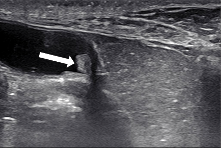
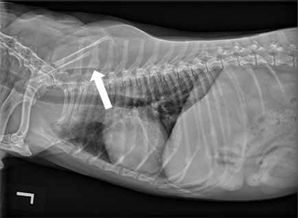
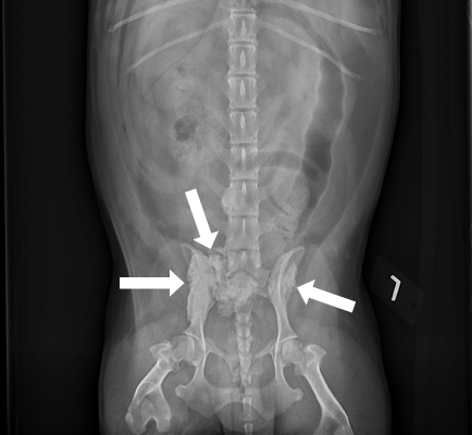
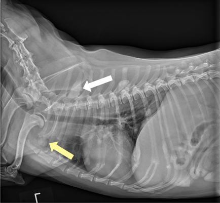
The CBC revealed leukocytosis of 21.8 K/µL (reference range, 6 to 17 K/µL), composed mostly of neutrophilia (19.6 K/µL [reference range, 3 to 12 K/µL]). Chronic inflammation caused by the UTIs is a potential reason for the neutrophilia present on Lily’s lab work. The serum biochemical profile revealed elevated alkaline phosphatase of 258 U/L (reference range, 20 to 157 U/L), which was potentially caused by the periosteal reactions of the bone. Radiography revealed multiple lung nodules and bone lesions in both the ilium and sacrum (FIGURES 2 AND 3), and abdominal ultrasonography showed urethral thickening, enlarged medial iliac lymph nodes, and a hepatic nodule (FIGURE 4).
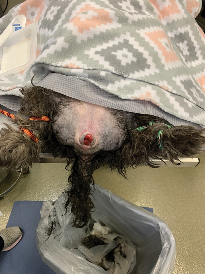
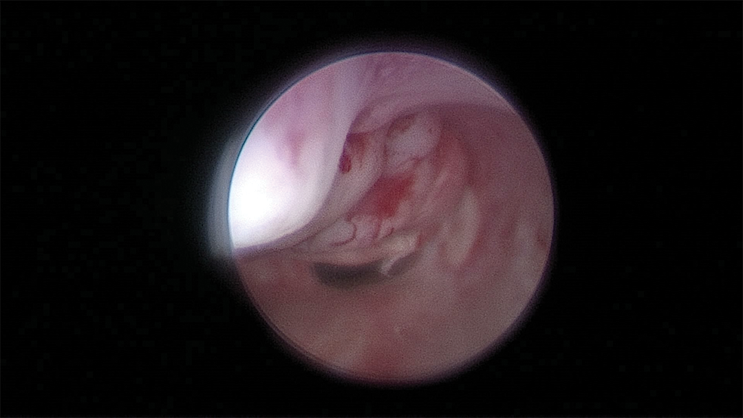
Lily was anesthetized for a cystoscopy, and during prep, it was noted that her mass was extending out of the urethra into the vulva (FIGURE 5). Multiple biopsy samples were taken from the urethra, and fine-needle aspiration was performed on the right wing of the ilium. Cytologic samples of the urethra and ilium both contained carcinoma cells. Histopathology definitively diagnosed high-grade iUC. Urine was collected through the scope for a urinalysis and urine culture. Cystocentesis is not recommended in patients with iUC due to seeding of the tumor to the abdominal wall. Urine samples should be collected via urinary catheterization or via a midstream, clean-catch urine sample into a sterile container.
Treatment Plan
After the diagnostic workup, an intravenous catheter was placed, and Lily was set up in the intensive care unit. She was started on a 3-µg/kg/h constant-rate infusion (CRI) of fentanyl overnight. A 0.1-mg/kg infusion of zoledronic acid was also administered. Zoledronic acid is a bisphosphonate used for osteoporosis in humans; however, in veterinary medicine, it is administered for bone pain and hypercalcemia. The drug works by inhibiting osteoclasts, thus halting the destruction of the bone and relieving bone pain. The following morning, there was no change in Lily’s CSU pain score; she was still at a 3 out of 4 and appeared to have windup present. Lily would vocalize when her kennel was approached, and without any manipulation. The fentanyl CRI was increased to 4 µg/kg/h, and she was started on a maintenance rate of a balanced crystalloid.
Chemotherapy was started immediately on Lily due to her large tumor burden, in hopes of decreasing the tumor burden and to help with her pain. She was administered 250 mg/m2 IV of carboplatin. The plan was to discontinue the grapiprant and then start her on 0.3 mg/kg piroxicam after a 6-day washout period. Amantadine was prescribed at 2 mg/kg q24h, along with codeine sulfate at 2 mg/kg q8h, and gabapentin was continued.
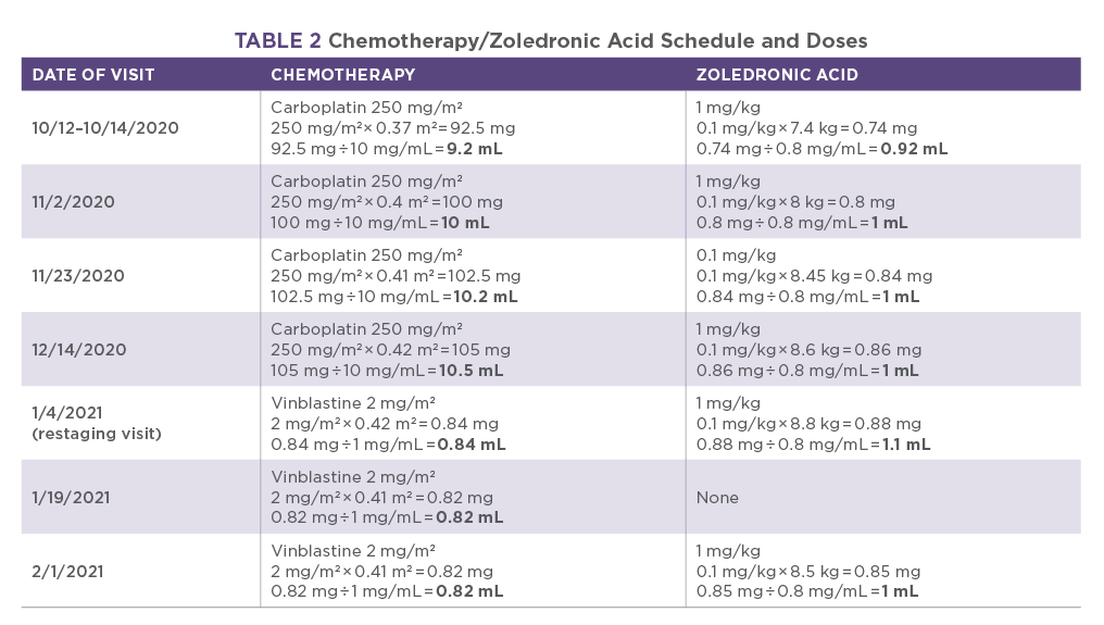
Lily received 3 doses of carboplatin and zoledronic acid at 3-week intervals (TABLE 2). It was reported on her second visit that she was more comfortable. She was allowing the owner to pick her up and was more sociable. Before her dose in January 2021, she was restaged to T3N2M1 (TABLE 1). It was noted that she was more uncomfortable recently, and the thought was that she was due for another dose of zoledronic acid. Restaging revealed progression of her disease, with advanced lung nodules and bone lesions and a new caudal abdominal mass (FIGURES 6 AND 7). Palliative care versus chemotherapy was discussed, and 2 mg/m2 IV vinblastine was administered along with zoledronic acid.
Outcome
Lily received a total of 3 doses of vinblastine. She tolerated the drug well with no side effects. However, on each visit, she started to become increasingly more painful. After her third dose, the owner chose to stop chemotherapy and continue palliative care at home. Lily was humanely euthanized with the referring veterinarian on February 17, 2021.
Discussion
Urinary bladder neoplasia accounts for approximately 1.5% to 2% of all canine malignancies.5 Around 90% of canine urinary bladder tumors are of epithelial origin, and the remaining 10% are of mesenchymal origin.5 iUC (epithelial malignancy) is the most common (90% to 95% of urinary bladder cancer) and is found to be an intermediate- to high-grade papillary infiltrative tumor in most canine patients.6 It is an aggressive form of cancer and is highly metastatic. It commonly affects the urinary bladder (most commonly the trigone), urethra, prostate, ureters, and kidneys. Metastasis at diagnosis is found in 20% of patients, and 50% have metastasis at necropsy. iUC commonly metastasizes to the lungs, regional lymph nodes, and distant lymph nodes. Less common sites of metastatic disease include the liver, spleen, and bone.2
The etiology of iUC in the canine patient is multifactorial. There are studies that show an association between iUC development and exposure to topical insecticides and herbicides.5 Other risk factors include obesity, female sex (around a 2:1 ratio of females to males), being spayed or neutered, administration of cyclophosphamide (due to cystitis and chronic inflammation), urinary calculi (chronic inflammation), and hereditary factors (Scottish terriers have an 18-fold increase).5 Shetland sheepdogs, beagles, wire fox terriers, and West Highland white terriers have a 3 to 5 times increased risk for iUC compared to mixed-breed dogs.5
iUC has a predilection for the trigonal region of the urinary bladder, where the ureters enter and where the urethra connects the urinary bladder to the outside of the body. If the mass becomes large enough and/or is in a critical location, it can block the ureters and/or urethra, causing urinary obstruction.
iUC is often not curable but is a cancer that can be well-controlled, allowing dogs to live with a good quality of life. However, the prognosis for iUC depends on several factors, with the following indicating a poorer prognosis:
- Patients with urethral involvement, due to the potential of obstruction and higher metastatic rate
- A higher TNM stage (TABLE 1)
- Prostatic involvement
- A younger age at diagnosis (the average age at diagnosis is 8 to 11 years7)
Lily was middle-aged at diagnosis (8.6 years), and her initial TNM stage was T3N1M1. Her higher TNM score was due to involvement of the vaginal vault (T3), regional lymph node metastasis (N1), and bone and lung metastasis (M1). Due to a higher TNM stage and her poor performance status, Lily was given a poor prognosis, with a survival time estimated at 1 to 3 months.
The standard of care for most dogs with iUC is focused on medical therapy, most commonly NSAIDs alone or NSAIDs combined with chemotherapy.1 One study showed that piroxicam induced remission in 20% of dogs and was associated with a median survival time of 195 days.5 Commonly used chemotherapy drugs for iUC are carboplatin, mitoxantrone, and vinblastine. Carboplatin is a synthetic platinum drug that works by producing cross-linkage of DNA, which modifies the DNA structure and inhibits DNA synthesis. Carboplatin combined with piroxicam is often utilized and has a remission rate of 38%.8 The oncologist in Lily’s case chose to start her on carboplatin and piroxicam due to her bone metastasis. In the clinical experience of this oncologist, carboplatin had been the most effective chemotherapy in reducing the pain from metastatic iUC. The other chemotherapy Lily received was vinblastine, which is a vinca alkaloid. Vinca alkaloids work by mitotic inhibition, which causes cell cycle arrest during metaphase. Vinblastine, administered without an NSAID, has been shown to induce a partial remission in 36% of dogs and stable disease in 50% of dogs.8 When combined with piroxicam, the median survival time was 299 days.8
Treatment is key to quality of life and survival time of patients with iUC. Those whose owners do not pursue treatment typically only survive a few weeks to a few months. The median survival time in dogs that do receive treatment can range from 200 to 300 days and longer in dogs taking advantage of more than 1 sequential treatment protocol.5
- Look for pollakiuria, stranguria, and hematuria as common clinical signs associated with urinary tract infection.
- Further investigation is recommended when a patient has recurrent urinary tract infections, with little to no response to antibiotic therapy.
- Cystocentesis is not recommended in a patient that is a high-risk breed or if a urinary bladder mass is suspected, due to potential seeding of the tumor to the abdominal wall.
References
- Fulkerson CM, Knapp DW. Tumors of the urinary system. In: Vail M, Thamm DH, Liptak JM, eds. Withrow and MacEwan’s Small Animal Clinical Oncology. 6th ed. Elsevier; 2020:645-656.
- Charney VA, Miller MA, Heng HG, Weng HY, Knapp DW. Skeletal metastasis of canine urothelial carcinoma: pathologic and computed tomographic features. Vet Pathol. 2017;54(3):380-386. doi:10.1177/0300985816677152
- Owen LN. TNM Classification of Tumours in Domestic Animals. World Health Organization (WHO). 1980. Accessed July 29, 2026. https://iris.who.int/server/api/core/bitstreams/a2994a51-7070-43e9-a7f9-f5338ac0e85e/content
- Decker B, Parker HG, Dhawan D, et al. Homologous mutation to human BRAF V600E is common in naturally occurring canine bladder cancer, evidence for a relevant model system and urine-based diagnostic test. Mol Cancer Res. 2015;13(6):993-1002. doi:10.1158/1541-7786.MCR-14-0689
- Knapp DW, Ramos-Vara JA, Moore GE, Dhawan D, Bonney PL, Young KE. Urinary bladder cancer in dogs, a naturally occurring model for cancer biology and drug development. ILAR J. 2014;55(1):100-118. doi:10.1093/ilar/ilu018
- Meuten DJ, Meuten TLK. Tumors of the urinary system. In: Meuten DJ, ed. Tumors in Domestic Animals. 5th ed. Wiley-Blackwell; 2017:656-675.
- Sommer BC, Dhawan D, Ratliff TL, Knapp DW. Naturally-occurring canine invasive urothelial carcinoma: a model for emerging therapies. Bladder Cancer. 2018;4(2):149-159. doi:10.3233/BLC-170145
- Arnold EJ, Childress MO, Fourez LM, et al. Clinical trial of vinblastine in dogs with transitional cell carcinoma of the urinary bladder. J Vet Intern Med. 2011;25(6):1385-1390. doi:10.1111/j.1939-1676.2011.00796.x