

Heartworm disease (dirofilariasis) has been diagnosed in all 50 states. It has been attributed to climate change, relocation of microfilariae-positive dogs, and prevalence of disease within wild canines. Currently, there are only 2 veterinary-approved approaches to management of heartworm-positive dogs: prevention and treatment of the adult worms. There are several medications approved by the U.S. FDA to prevent heartworm disease but only 1 approved adulticide: melarsomine. The benefits of treatment need to outweigh the risks, which, in this case, ended up being severe. The treatment needs to be administered deep into the epaxial muscles and can contribute to pain and myositis at the site of injection. There have been 2 reported cases of melarsomine causing such severe myositis that it led to neurologic deficits in the spinal cord and sepsis.
Take-Home Points
- The goal of adulticide therapy is to minimize any clinical condition the animal may be experiencing.
- Melarsomine is an arsenic drug and is the only drug approved by the U.S. FDA to kill adult heartworms.
- Patient comfort depends on observational and nursing skills of the supervising veterinary nurse/technician.
- Advanced disease may lead to complications such as systemic inflammatory response syndrome (SIRS) increasing the risk of sepsis, in which patients may become recumbent, requiring extensive nursing care and a vigilant nursing team to identify and respond to subtle abnormalities.
- SIRS can cause increased vascular permeability, leading to third spacing, a hypercoagulable state, and ultimately death.
Clinical heartworm disease (dirofilariasis) is observed in the late stage of infection by the parasite Dirofilaria immitis within dogs. Melarsomine is currently the only arsenical veterinary drug approved to treat heartworm disease; its efficacy rate is 92% to 98%.1 The goal of therapy is to kill adult D immitis worms while not harming the patient.
The approved method of administration is an injection deep within the epaxial muscles between L3 and L5 in 3 separate doses. A single injection is administered and then repeated in 2 doses a month later (24 hours apart). If the patient is discharged the same day of treatment, the owner is instructed to monitor for any signs of anaphylaxis such as vomiting, ataxia, fever, inappetence, and lethargy.1
It is typical for the injection sites to feel warm to the touch and develop a lump that fades away. Because the treatment needs to be administered deep into the epaxial muscles, it can contribute to pain and myositis at the site of injection. Two reported cases of melarsomine injection caused such severe myositis that it led to neurologic deficits in the spinal cord and sepsis.2
Other adjunctive therapies exist:
- Doxycycline is used to eliminate Wolbachia species bacteria, which are necessary for the D immitis microfilaria to survive and become adult worms.
- Macrocyclic lactones such as ivermectin can also be beneficial to kill larval stages and prevent new infections during treatment.3
Pet owners must also restrict their pet from exercising during treatment to minimize the risk for a fatal pulmonary thromboembolism as the adult worms die.
History and Presentation
Sophie, a 9-year-old spayed female Brittany spaniel, had tested positive for Anaplasma platys and heartworm disease in November 2023. She had begun treatment by her primary veterinarian for heartworm disease with melarsomine on February 5, 2024. The first dose of melarsomine (73 mg IM) was administered in the left epaxial muscle; the second dose (75 mg) was administered 30 days later in the right epaxial muscle and a third dose (73 mg) administered in the left epaxial muscle the next day, for a total of 3 doses.
Sophie was presented to the emergency service on August 26, 2024, due to difficulty standing and for evaluation of alopecia bilaterally on her dorsal lumbar region. She was also experiencing episodes of urinary accidents in the home and had a decreased appetite for approximately 2 days before presentation.
Initial Assessments and Diagnostics
On initial presentation, Sophie was febrile with a temperature of 40 °C (104 °F) (reference range, 37.7 to 39.3 °C [100 to 102.9 °F]).4 She was tachycardic and panting with a heart rate of 180 beats per minute (bpm) (reference range, 80 to 120 bpm) and a systolic blood pressure of 110 mm Hg measured via Doppler ultrasonography.
She was ambulatory on all 4 limbs but had dermal ulceration and oozing along her lumbar epaxial muscles bilaterally (FIGURE 1). Her mucous membranes were noted to be icteric.
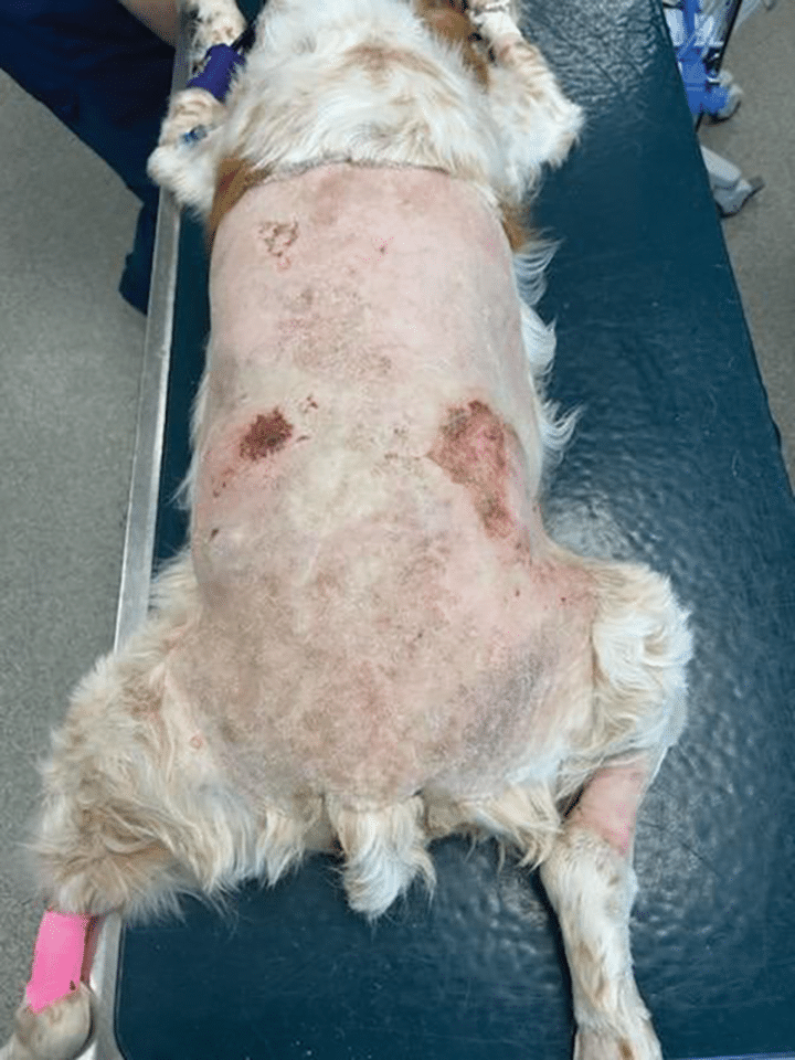
Figure 1. Dermal ulceration on presentation.
Blood was obtained for a serum biochemical profile, a coagulation profile, urinalysis, and an IDEXX SNAP 4Dx PLUS enzyme-linked immunosorbent assay (ELISA) test. The ELISA test was positive for Anaplasmosis and heartworm disease.
A CBC revealed a leukocytosis of 49.62 K/µL (reference range, 5.05 to 16.76 K/µL) with a neutrophilia of 42.7 K/µL (reference range, 2.95 to 11.64 K/µL) and an elevated prothrombin time of 137 seconds (reference range, 72 to 102 seconds). Serum biochemical profile revealed a blood glucose of 35 mg/dL (reference range, 70 to 143 mg/dL), albumin of 2 g/dL (reference range, 2.2 to 3.9 g/dL), alanine aminotransferase of 148 U/L (reference range, 10 to 125 U/L), and alkaline phosphatase 1827 U/L (reference range, 23 to 212 U/L).
Thoracic radiography yielded no clinical abnormalities. Abdominal ultrasonography showed chronic vacuolar inflammatory hepatopathy and possible inflammation within the aorta, along with enlarged inguinal and iliac lymph nodes. These findings were suggestive of inflammation and early thrombosis within the aorta and liver.
Based on Sophie’s clinical presentation, she had already met the criteria indicating that she was experiencing systemic inflammatory response syndrome (SIRS) (BOX 1). It has been found that typically SIRS leads to sepsis; however, SIRS can manifest itself in the absence of infection and mimic the clinical course of sepsis.5
- Leukocytosis or leukopenia
- Hypothermia or hyperthermia
- Tachycardia
- Tachypnea
SIRS = systemic inflammatory response syndrome
Treatment
Sophie was subsequently admitted to the intensive care unit. An 18-g IV catheter was placed into the right cephalic vein, and a crystalloid bolus of 10 mL/kg was administered intravenously over 30 minutes. She was then started on crystalloids with 5% dextrose at a rate of 3.4 mL/kg/h.
Additional blood samples were obtained to send out for a fever-of–unknown origin panel to the reference laboratory. Ampicillin-sulbactam was started at 43 mg/kg IV q6h, along with enrofloxacin at 10 mg/kg q24h. Sophie was started on a fentanyl constant-rate infusion at 3 µg/kg/h. A peripherally inserted central catheter was placed to allow for serial blood analysis monitoring.
On the second day of hospitalization, the veterinarian and owners elected to anesthetize Sophie and have a board-certified surgeon debride the bilateral epaxial wounds. The musculature underneath the adipose tissue was green/brown in color and did not have any visible perfusion. Debridement was continued until grossly normal tissue was identified (FIGURES 2 AND 3).
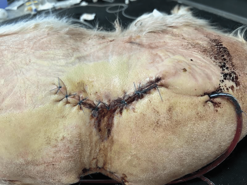
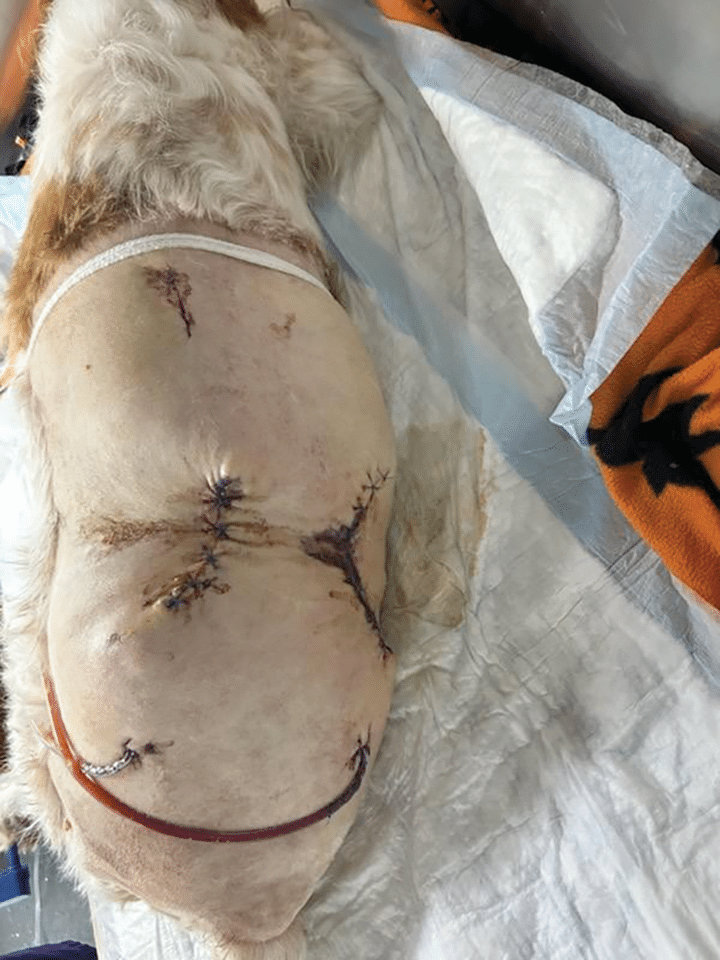
Upon anesthetic recovery, a 5.5-Fr × 13-cm central line was placed as well as an 8-Fr nasogastric tube to allow for enteral nutrition. When lab work was rechecked, her albumin had decreased from 2 g/dL to 1.6 g/dL. She received a fresh frozen plasma (FFP) transfusion of 10 mL/kg over 4 hours.
A cytologic evaluation of the necrotic tissue that was debrided from her left lumbar epaxial region yielded fibrinous material with occasional degenerate neutrophils and intracellular cocci bacteria; however, a culture yielded no growth on multiple samples of the necrotic tissue.
Nursing care was extensive, with temperature, heart rate, respiration, pain evaluation, and nutritional assessment, along with blood glucose and Doppler ultrasonography blood pressure checks every 4 hours to ensure Sophie would not start trending toward sepsis.
Approximately 9 hours postoperatively, the veterinarian elected to administer a second FFP transfusion of 10 mL/kg over 4 hours given that her recheck lab work yielded a continued deficiency in her albumin of 1.7 g/dL and an elevation of her bilirubin of 4 mg/dL. Grossly, her mucous membranes were more icteric, in addition to a new finding of ventral abdominal pitting edema. She continued to be inappetent, and a low-fat diet was started at 324 kcal/day via the nasogastric tube.
On the third day of hospitalization, the pitting edema and icterus began to worsen. Sophie was persistently tachycardic at 140 bpm, and recheck lab work yielded a worsening leukocytosis of 77 K/µL, neutrophilia of 67 K/µL, a nonregenerative anemia of 13% (reference range, 35% to 45%), an elevated partial thromboplastin time of 137 seconds (reference range, 72 to 102 seconds), a continuously increasing total bilirubin of 6.4 mg/dL (reference range, 0 to 0.9 mg/dL), and a creatinine that steadily increased to 1.5 mg/dL (reference range, 0.5 to 1.8 mg/dL). An increased respiratory rate was observed, and point-of-care ultrasonography (POCUS) revealed a scant amount of bilateral pleural effusion, which was not significant enough for collection. She received a third 10-mL/kg FFP transfusion administration over 4 hours.
Sophie’s nursing care became more intensive with hourly respiratory checks, frequent POCUS scans, and recumbency care added to her already extensive nursing care as she was increasingly becoming nonambulatory. The continued drop in albumin and development of pleural effusion support the indication of continued treatment for SIRS. The new lab work indicated that she was experiencing multiple organ dysfunction syndrome (MODS), which is defined by 2 or more dysfunctional organs in patients with SIRS.6 Sophie was experiencing the following dysfunctions:
- Coagulation by having increased clotting times
- Hepatobiliary dysfunction by the continuous increase in total bilirubin
- Kidney dysfunction by a steadily increasing creatinine
- Vascular dysfunction indicated by her third spacing and vascular leak
- Endocrine dysfunction by persistent hypoglycemia in the absence of dextrose supplementation
On the fourth day of hospitalization, she was crossmatched and received a packed red blood cell transfusion of 10 mL/kg over 4 hours in response to her anemia, which was well-tolerated. She remained nonambulatory, and a 10-Fr Foley urinary catheter was placed to monitor urine production. A repeated POCUS scan revealed static pleural effusion and no peritoneal effusion.
Outcome
The evening of her fourth day, she had become ambulatory with assistance. After her treatments, walking her outside was attempted. On the way back in, she collapsed and experienced cardiac arrest. Closed-chest resuscitation was initiated per RECOVER (Reassessment Campaign on Veterinary Resuscitation) Initiative guidelines7 and continued for 16 minutes until her owners elected to discontinue.
- Maintain veterinary nurse/technician-to-patient continuity in the intensive care unit to help detect subtle changes in a patient’s status.
- Emphasize the importance of knowing the normal limits of patients’ vital signs to recognize abnormalities quickly.
- Be familiar with the patient’s underlying condition and the disease processes that could lead to complications.
Summary
The use of melarsomine to treat heartworm disease in patients requires a careful assessment of risks versus benefits. Given the adulticide’s high efficacy, the decision to proceed with treatment was well-supported. Unfortunately, in this case, when Sophie had presented to the emergency service, she was already experiencing SIRS, which has a mortality rate higher than 40%.8 Throughout the course of treatment, she developed comorbidities that developed into MODS, from which she was ultimately not able to recover.
References
- Nelson T, McCall J, Moorhead A, Starkey L, Ames M. Highlights of the Canine Guidelines for the Prevention, Diagnosis, and Management of Heartworm Infection in Dogs. American Heartworm Society. Revised 2024. Accessed March 30, 2026. https://d3ft8sckhnqim2.cloudfront.net/images/AHS_Canine_Guidelines_Highlightsweb31JUL2024.pdf
- Arnold SA, Kent M, Ruby JL, Clarke LL, Barber RM, Platt SR. Extensive epidural steatitis and epaxial myositis associated with intramuscular injection of melarsomine dihydrochloride in two dogs. VCOT Open. 2020;03(01):e46-e51. doi:10.1055/s-0040-1710070
- Cole TB, Lynn RC. Drugs for the treatment of helminth infections: anthelmintics. In: Boothe DM, ed. Small Animal Clinical Pharmacology & Therapeutics. 2nd ed. Elsevier; 2012:451-465.
- Campbell MT. Hemolymphatic, immunologic, and oncology emergencies. In: Norkus C, ed. Veterinary Technician’s Manual for Small Animal Emergency and Critical Care. Wiley-Blackwell; 2012:221-261.
- Goggs RAN, Lewis DH. Multiple organ dysfunction syndrome. In: Silverstein D, Hopper K, eds. Small Animal Critical Care Medicine. Saunders; 2023:35-46.
- Sharp CR. Systemic inflammatory response syndrome, sepsis, and multiple organ dysfunction syndrome. In: Drobatz KJ, Hopper K, Rozanski E, Silverstein DC, eds. Textbook of Small Animal Emergency Medicine. Wiley-Blackwell; 2019:1030-1037.
- Hopper K, Epstein SE, Burkitt-Creedon JM, et al. 2024 RECOVER guidelines: basic life support. Evidence and knowledge gap analysis with treatment recommendations for small animal CPR. J Vet Emerg Crit Care (San Antonio). 2024;34(suppl 1):16-43. doi:10.1111/vec.13387
- Waxman C. Recognizing, treating, and monitoring systemic inflammatory response syndrome and sepsis. Todays Vet Nurse. 2020;3(2):35-43.