
To take the CE quiz, click here.
This quiz is open until August 2028.
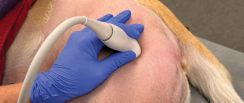
Locoregional anesthesia can be a helpful component of a balanced anesthesia plan for a veterinary patient. However, poor training accessibility makes it an often underutilized component. This article serves as a resource for veterinary nurses regarding the pharmacology of local anesthetics and their side effects as well as clinical applications of abdominal and thoracic locoregional anesthesia with a step-by-step guide for several ultrasound-guided nerve blocks.
Take-Home Points
- Local anesthetic agents block pain signaling for various durations dependent on the agent and adjuvant.
- Local anesthetic systemic toxicity occurs from cumulative doses; thus, the total dose should be carefully calculated.
- Ultrasound-guided nerve blocks require a basic understanding of ultrasound techniques and the ability to perform accurate in-plane needle guidance.
- Fascial plane blocks enable multiple nerves to be blocked between muscle planes and are often used for locoregional anesthesia of the abdomen and thorax.
- Intercostal nerve blocks are shown to be more successful with ultrasound guidance.
Locoregional anesthesia is the global term used to describe locally administered anesthesia (e.g., line block) and regionally administered anesthesia at the level of specific nerves (e.g., femoral nerve block). Local anesthetics block transmission of painful stimuli through the central nervous system to provide a loss of pain sensation in a specific area of the body. When used appropriately, locoregional anesthesia improves pain control and decreases reliance on opioids and inhalant anesthetics—all of which lead to a safer anesthesia experience.1 Improving local pain control without reliance on centrally acting medications results in fewer side effects and shorter hospitalization times.2 Locoregional anesthesia is also shown to reduce central sensitization and wind-up pain.3 In addition, the supplies required for administration of locoregional anesthesia are widely available and cost effective. Although locoregional anesthesia may be performed under light sedation in humans, veterinary patient compliance and awareness may necessitate general anesthesia to accompany locoregional anesthesia. Nevertheless, locoregional anesthesia techniques are becoming more widely available and are gaining popularity as a component of a balanced analgesic plan for veterinary patients.
Basics of Nerves Using Ultrasonography
To perform an ultrasound-guided peripheral nerve block, the provider needs to have a basic understanding of ultrasonography and nerve block techniques. (The basics of ultrasonography are beyond the scope of this article, but readers can watch VIDEO 1 for a concise explanatory video.) The provider should be familiar with the available ultrasonography machine, including its depth capabilities, focus position, gain settings, and color Doppler. Many blocks discussed in this article require a shallow depth setting, which may be difficult to achieve with some ultrasonography machines.
The ideal ultrasonography unit will have a linear and/or small curvilinear probe with a very shallow depth setting (< 2 cm). Large curvilinear probes are not well-suited for nerve blocks. All nerve block techniques are considered in-plane, meaning the entire needle and needle tip should be visible on the screen at all times (FIGURE 1). This is the same technique used for ultrasound-guided cystocentesis.
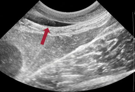
Figure 1. In-plane needle technique. Needle (arrow) is shown in plane with the ultrasound probe terminating in the injectate.
With ultrasonography, nerves generally appear as hollow, tubular structures with a hypoechoic to anechoic central region and hyperechoic rim. Some larger nerves may have an internal “honeycomb” appearance.4 Nerves can be differentiated from vasculature by color Doppler (FIGURE 2).
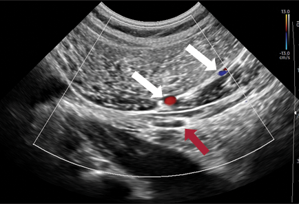
Figure 2. Sciatic nerve (red arrow) compared to nearby vasculature with color Doppler (white arrows).
The long-standing theory has been that only extraneural (outside the nerve) blocks should be performed due to the risk for nerve damage during direct injection. Although the literature is currently divided on this theory, extraneural injections are effective at producing nerve blockade and remain the safest option.5 During injection, the goal is to place the injectate in close proximity to the nerve, and potentially circumferentially for optimal blockade, but not within the nerve itself.
Local Anesthetics
Mechanism of Action
Local anesthetic agents provide nociceptive blocking effects by binding to sodium channels within nerve cells. The local anesthetic diffuses through the cell membrane in its inactive, nonionized form. Within the cell, it ionizes and binds to sodium channels. Then, the local anesthetic inhibits the movement of sodium into the cell by creating an electrostatic field that repels positively charged ions such as sodium.6 Because cells rely on the movement of sodium ions to initiate cell communication (i.e., action potential propagation), the interruption of this pathway prevents peripheral nerve cells from relaying signals to the central nervous system, thus a blockade.
The primary goal of locoregional anesthesia is to provide nociceptive sensory blockade. A secondary goal is to reduce unwanted side effects such as motor blockade. At lower concentrations of local anesthetics, sensory blockade happens earlier than motor blockade. When motor blockade happens, motor function usually returns before sensory function.7 When locoregional anesthesia is appropriately administered, muscle tone is usually lost first, followed by the sensation of sharp pain, touch, and motor function. Warmth and deep pain/pressure are the final sensory losses; thus, even with adequate nerve blockade, pain from cautery is unlikely to be blocked.8,9
There are 3 primary factors in the formulations of local anesthetics that create clinical differences.10
- Amount of nonionized drug within the body: Only the nonionized form of a local anesthetic can diffuse into a cell. The more nonionized drug, the more rapid the onset of blockade. Ionization is affected by pH, and this value may be described as pKa, or the pH at which 50% of the local anesthetic is nonionized.10 In general, the higher the pKa, the less drug exists as nonionized at the body’s pH and the slower the onset.
- Degree of protein binding: Drugs with a higher degree of protein binding have a longer duration of action.10
- Degree of lipid solubility: Greater lipid solubility increases drug diffusion into the cell and thus potency.10
Toxicity
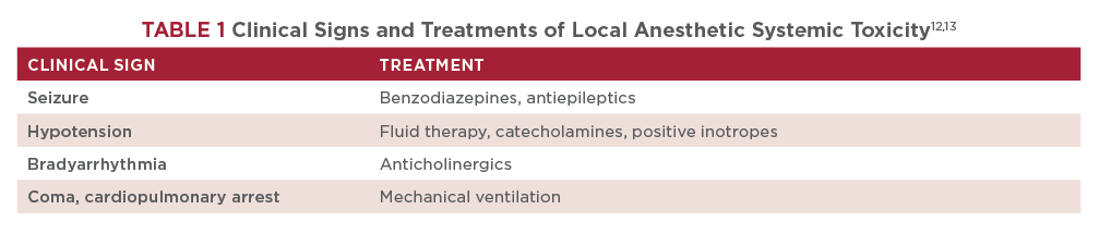
Local anesthetics may be systemically absorbed through either accidental intravascular injection or injection site absorption.11 Local anesthetic systemic toxicity (LAST) leads to negative neurologic and cardiovascular effects, including altered mental state, seizures, coma, hypotension, bradyarrhythmia, and cardiac arrest.12 Cats are particularly sensitive to the cardiotoxic effects of local anesthetics and, thus, have lower maximum doses.13 Local anesthetic toxic effects are cumulative; multiple dosage sites should be calculated and divided from a single total dose (Box 1).
Total injections: 4
Volume per injection: 0.2 mL/kg
Local anesthetic: Bupivacaine 0.5% (concentration 5 mg/mL)
Dose: 2 mg/kg
Patient weight: 17 kg (37.5 lb)
Total local anesthetic volume: ([dose × patient weight in kg] / concentration) = 6.8 mL
Total volume per injection: volume per injection × patient weight in kg = 3.4 mL
Local anesthetic volume per injection: total local anesthetic volume / number of sites = 1.7 mL
Diluent volume per injection: total volume per injection − local anesthetic volume per injection = 1.7 mL
For this patient, 4 syringes are predrawn. Each syringe contains 1.7 mL of saline mixed with 1.7 mL of bupivacaine.
Example 2
Total injections: 6
Volume per injection: 0.05 mL/kg
Local anesthetic: Ropivacaine 0.75% (concentration 7.5 mg/mL)
Adjuvant: Buprenorphine (concentration
0.3 mg/mL, dose 0.003 mg/kg)
Dose: 1 mg/kg
Patient weight: 3.2 kg (7 lb)
Total volume per injection: volume per injection × patient weight in kg = 0.16 mL
Total local anesthetic volume: ([dose x patient weight in kg] / concentration) = 0.42 mL
Total adjuvant volume: ([dose x patient weight in kg] / concentration) = 0.03 mL
Total diluent volume: (volume per injection x number of injections) – (local anesthetic total volume + adjuvant total volume) = 0.51 mL
For this patient, 0.16 mL of ropivacaine is mixed into a sterile solution with 0.03 mL buprenorphine and 0.51 mL saline. Six syringes are drawn from the solution with 0.16 mL in each syringe.
Note: If there is a minimum volume per site that is greater than the prescribed volume per injection, more diluent may need to be added; however, block efficacy and duration may be decreased.
For patients with suspected or known LAST, aggressive supportive care should be initiated (TABLE 1). Lipid emulsion may be beneficial for patients with known accidental IV administration.13
Drug Combinations
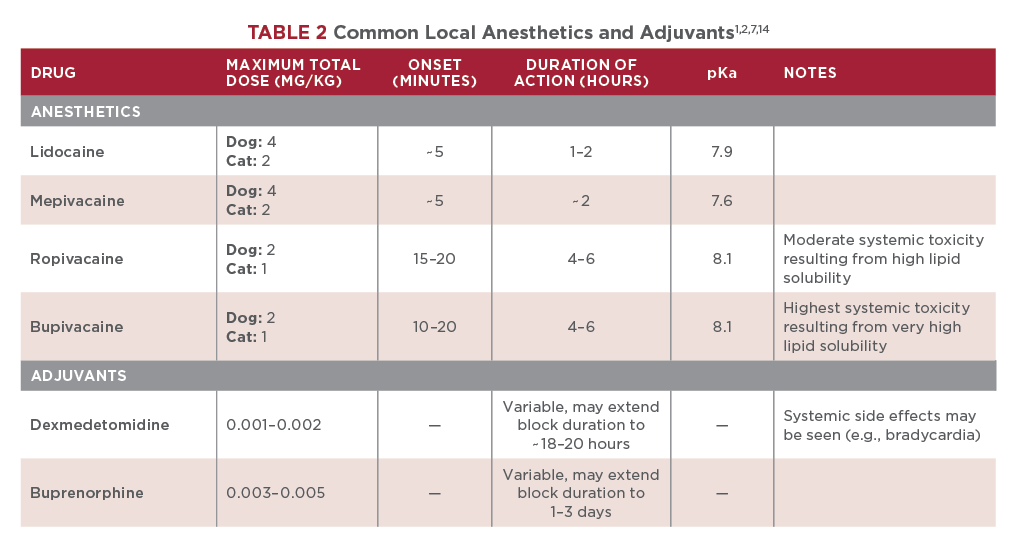
Local anesthetics may be combined with various adjuvants, such as NSAIDs, ketamine, steroids, or other opioids, to extend their duration of action and potency (TABLE 2). Other adjuvants may be used as well, but research has shown variable results. Therefore, this article will not focus on other adjuvants. Of note, combining multiple local anesthetics typically results in decreased potency and duration of action and is not recommended.1,14
Dexmedetomidine
Dexmedetomidine, an α₂ agonist, may be combined with a local anesthetic to prolong the duration of action by inducing local vasoconstriction. The results vary, but some combinations with a long-acting local anesthetic have increased the duration of action by an additional 12 hours.1 Depending on the area of injection and dose, systemic side effects (e.g., bradycardia) may be observed. The dose should be adjusted accordingly.14
Buprenorphine
Although morphine is the most commonly used opioid for spinal anesthesia, buprenorphine, a partial opioid receptor µ-agonist/κ-antagonist, has pharmacologic effects that make it a more effective local anesthetic adjuvant for peripheral nerve blocks. Buprenorphine binds and blocks sodium channels in the cell membrane with a higher affinity than most local anesthetics, causing similar effects as local anesthetics and demonstrating a slow onset and long duration of action.1,14,15 Studies have shown that the addition of buprenorphine to peripheral nerve blocks of the brachial plexus can extend analgesia to 30 to 96 hours.1,15
Dilutions
Local anesthetics are commonly diluted with sterile saline or sterile water to increase the volume. Mixing a local anesthetic with saline in a 1:1 ratio has been shown to increase the volume without compromising efficacy.1,14 Blocks will list a recommended volume per injection site, which is often greater than the volume of local anesthetic. To achieve the correct volume, many blocks require dilution. The maximum total dose is based on cumulative doses and is not a per site dose (e.g., total dose of 2 mg/kg divided by the number of sites, not 2 mg/kg per site).
Preparation
Patients should be heavily sedated or anesthetized and placed in the appropriate position (as described for each specific block below). The surrounding hair should be clipped, and the skin should be aseptically prepared. Isopropyl alcohol is typically used to improve the contact of the probe to the patient’s skin. For damaged tissues, sterile lubricant can be used instead of isopropyl alcohol. Supplies should be prepared ahead of time, and these include:
- Drugs: Drugs should be drawn into individual doses and well-labeled to prevent accidental intravenous administration.
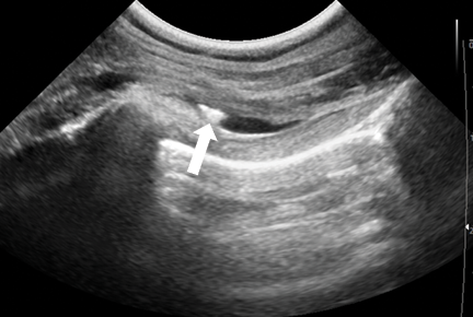
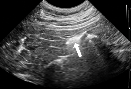
- Needles: Needle size can be determined by ultrasonography measurement. Most blocks require a 22-gauge 1.5-in needle. Small or thin patients may require a shorter 25-gauge needle, and large-breed dogs may require a longer spinal needle (e.g., 2.5 in to 3 in). Air should be removed from all needles by drug priming. Because air reflects ultrasound waves, accidental administration of air into the block region not only will make visualization more difficult but may also impede dispersion of the local anesthetic (FIGURE 3). All syringes should have a new needle placed before injecting to ensure the sharpest injection point and decrease the risk for needle contamination.
- Optional T-port assistance: For those with less experience performing ultrasound-guided nerve blocks, a T-port on the end of the needle and an assistant, who controls aspiration and drug administration, can help steady the needle and provide more accurate drug delivery (FIGURE 4).
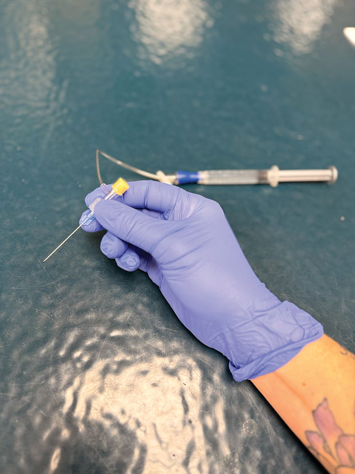
FIGURE 4. Optional T-port for assistance. Entire system, including needle, must be primed to remove air.
Block Techniques
Abdomen
Nerve innervation of the abdomen in dogs and cats can vary. In general, the lateral abdominal walls and cranial abdomen are innervated by spinal nerves T6 through T10, and the remainder of the abdomen is innervated by spinal nerves T11 through L3.16-20 These spinal nerves branch from the spinal cord and traverse through muscle groups to provide cutaneous sensory information. These nerves can be blocked as they dip between muscle groups by fascial plane blocks. Fascial plane blocks use the space between muscle groups to deposit large volumes of local anesthetics (usually diluted to achieve desired volume) and block several nerves at once. (Providers should always double-check the maximum dose of a local anesthetic before administration to prevent iatrogenic LAST.) Fascial plane blocks are performed based on anatomic landmarks, unlike most orthopedic blocks, which are commonly performed by viewing the specific nerves. Because of the high number and complex anatomy of nerves that innervate the abdomen, fascial plane blocks are used for abdominal locoregional anesthesia.
Transversus Abdominis Plane (TAP) Block
Relevant Anatomy
The lateral abdominal wall is composed of 3 muscle layers. The most superficial layer is the external oblique; moving deeper, the internal oblique layer is followed by the deepest layer, the transversus abdominis. The cranial abdomen, near the midline of a patient, is composed of different muscle groups. The internal oblique ends and is replaced by the rectus abdominis muscle between the external oblique and transversus abdominis.
Performing a Lateral TAP Block
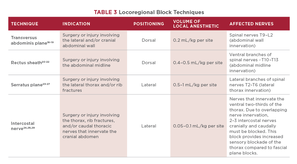
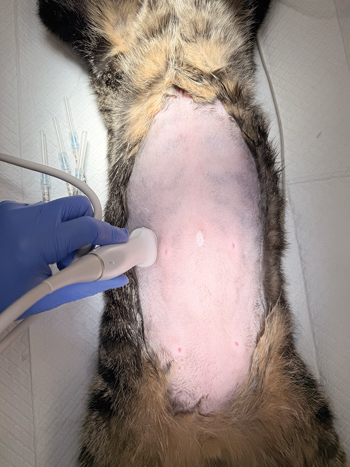
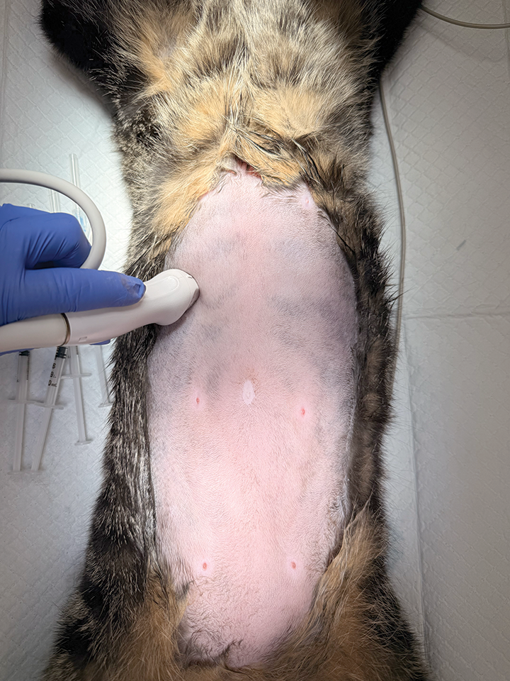
A lateral TAP block aims to dispense a high volume of local anesthetic solution into the fascial plane above the transversus abdominis and below the internal oblique (TABLE 3). The ultrasound probe is placed on the lateral abdomen in a sagittal orientation at approximately the level of the umbilicus (FIGURE 5A). The TAP is identified between the internal oblique and transversus abdominis, and the needle is placed within the TAP (FIGURE 6). After aspirating to confirm the needle is not within vasculature, a small test injection may be performed to evaluate appropriate spread of the anesthetic solution within the fascial plane, called hydrodissection. The full volume of injectate can be administered when appropriate hydrodissection is confirmed. This procedure is then repeated on the opposite side.
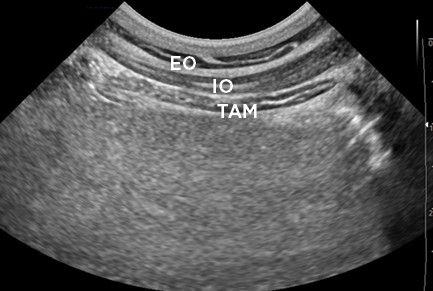
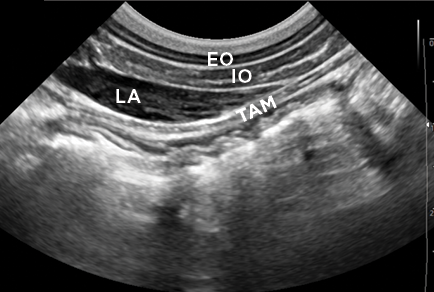
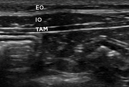
EO = external oblique
IO = internal oblique
LA = local anesthetic
TAM = transversus abdominis muscle
Performing a Subcostal TAP Block
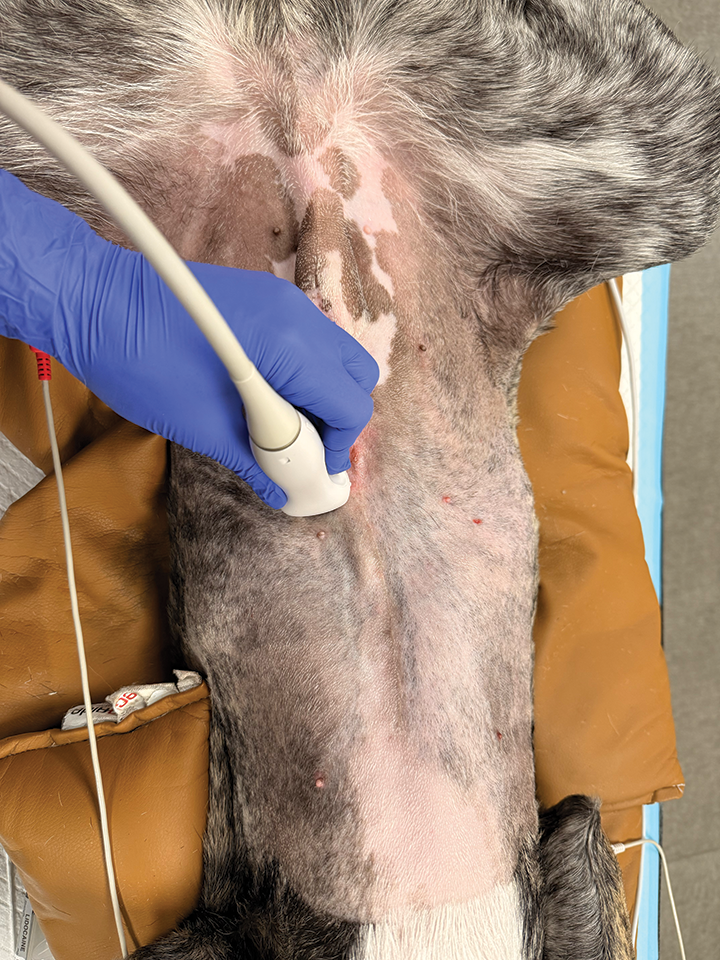
A subcostal TAP block aims to dispense a high volume of local anesthetic solution into the fascial plane above the transversus abdominis and below the rectus abdominis (TABLE 3). The ultrasound probe is placed near the level of the costochondral junction, caudal and parallel to the last rib, in a diagonal orientation (FIGURE 5B). The probe is moved medially until the TAP is identified between the rectus abdominis and transversus abdominis, and the needle is placed within the TAP (FIGURE 7). Injection continues as described above and repeated on the opposite side.
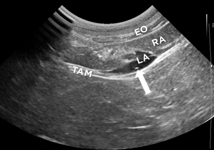
EO = external oblique
LA = local anesthetic
RA = rectus abdominis
TAM = transversus abdominis muscle
Rectus Sheath Block
Relevant Anatomy
The muscles along the cranial abdominal midline include the rectus abdominis, which is above the transversus abdominis. The rectus abdominis is enclosed by a fibrous tunnel called the rectus sheath. The rectus sheath is divided into an external part that is above the rectus abdominis and an internal part that is below the rectus abdominis.
Performing a Rectus Sheath Block
The ultrasound probe is first placed over the midline, cranial to the umbilicus, in a transverse orientation (FIGURE 5C). The probe is moved laterally to observe the lateral edge of the rectus abdominis and medial edge of the transversus abdominis. Note that the degree of overlap can vary among patients (i.e., the transversus abdominis may extend closer to or further from the midline). The internal rectus sheath, which is beneath the rectus abdominis and above the transversus abdominis, can be seen as a double, hyperechoic line (FIGURE 8). However, the double line may be difficult to visualize with some ultrasonography machines. The needle is advanced through the rectus abdominis, superficial to the internal rectus sheath. The double, hyperechoic line of the rectus sheath should be beneath the needle. Injection continues as described above and repeated on the opposite side (TABLE 3).
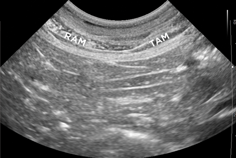
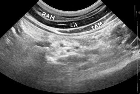
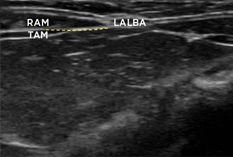
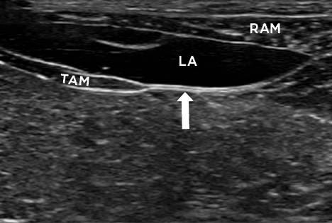
LA = local anesthetic
LALBA = linea alba
RAM = rectus abdominis muscle
TAM = transversus abdominis muscle
Thorax
The thorax is innervated by the thoracic spinal nerves T2 through T13.23,24,29 The spinal nerves branch from the spine and run along the caudal aspect of the ribs in a neurovascular bundle, which also contains the intercostal vein and artery. The intercostal nerves have several different branches, which provide cutaneous innervation to various parts of the thorax. Deviations from this anatomy include the spinal nerves T1 and T13.24,29 Spinal nerve T1 contributes to the brachial plexus and does not provide cutaneous sensory information from the thorax. Spinal nerve T13 branches to become the costoabdominal nerve.
Like the abdomen, locoregional anesthesia of the thorax relies heavily on fascial plane blocks with the technique varying according to the region requiring sensory blockade. However, the spinal nerves can also be blocked directly via intercostal nerve blocks performed at the caudal border of the rib associated with the desired thoracic spinal nerve. A risk for iatrogenic thoracic puncture, leading to pneumothorax, is present when performing thoracic blocks.
Serratus Plane Block
Relevant Anatomy
The craniolateral chest wall contains 3 muscles superficial to the ribs, which appear with acoustic shadowing. From superficial to deep, these muscles are the cutaneous trunci, latissimus dorsi, and serratus ventralis. The external intercostal muscles lie beneath the serratus ventralis. The superficial serratus ventralis fascial plane lies between the latissimus dorsi and serratus ventralis, and the deep serratus ventralis fascial plane lies between the serratus ventralis and external intercostal muscles. The superficial and deep approaches have been shown to provide analgesia in humans, but some reports from human medicine cite the deep approach as providing increased analgesia.26
Performing a Serratus Plane Block
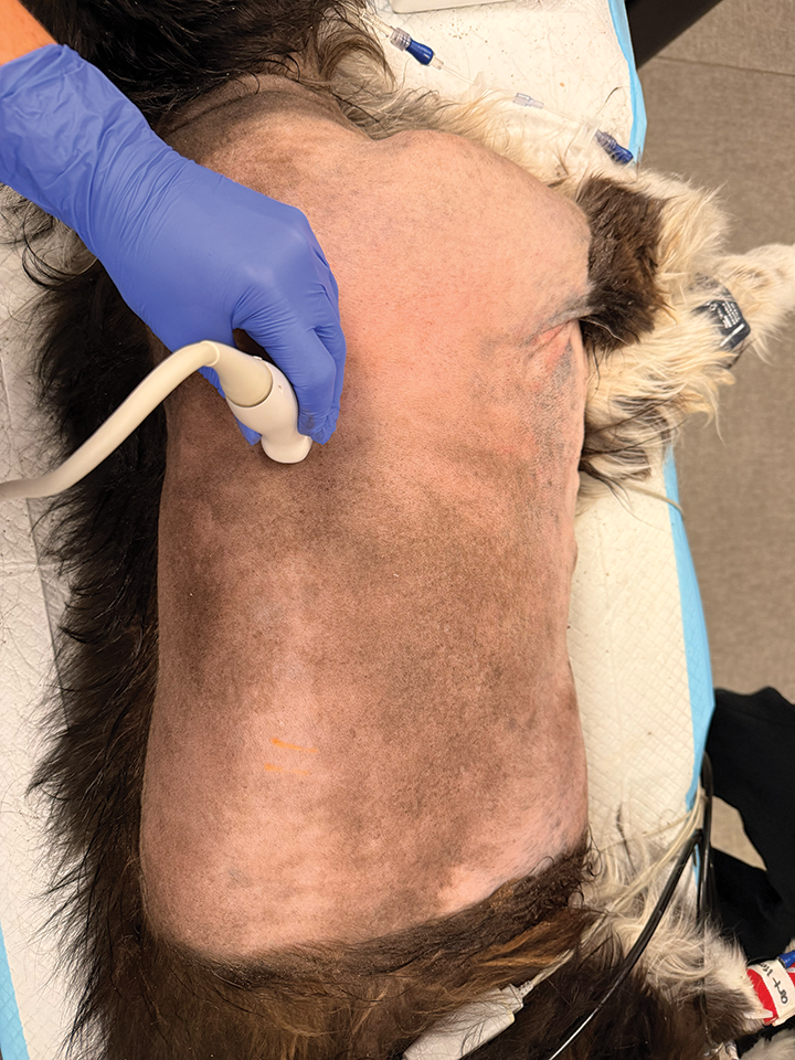
Extend the thoracic limb cranially. The ultrasound probe is placed caudal to the thoracic limb, over the lateral thorax, at the level of the 4th and 5th intercostal space in sagittal orientation (FIGURE 9). The needle is advanced into the desired (superficial or deep) serratus ventralis fascial plane (FIGURE 10). Injection continues as described above and is repeated at the 5th and 6th intercostal space (TABLE 3).
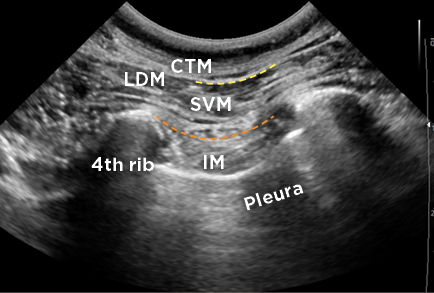
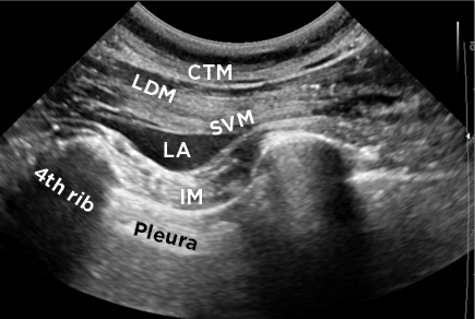
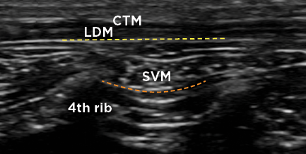
CTM = cutaneous trunci muscle
IM = intercostal muscles
LA = local anesthetic
LDM = latissimus dorsi muscle
SVM = serratus ventralis muscle
Intercostal Nerve Block
Relevant Anatomy
Each intercostal nerve runs along the caudal border of the rib in a neurovascular bundle. Within that bundle, an intercostal vein lies most cranially, followed by the intercostal artery, and then, most caudally, the intercostal nerve. The neurovascular bundle is surrounded by connective tissue and muscle. On ultrasonography, the ribs will be visualized with acoustic shadowing; the neurovascular bundle lies on the caudal border, superficial to the pleural surface of the thoracic wall, which is visualized as a hyperechoic line with distant air artifact from lung tissue (FIGURE 11).
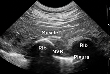
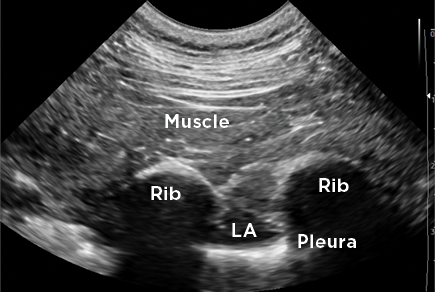
LA = local anesthetic
NVB = neurovascular bundle
Performing an Intercostal Nerve Block
Inexperienced providers will benefit from marking the rib spaces on the skin ahead of time to prevent rib counting errors. The ultrasound probe is positioned dorsally over the thorax with the ribs of the predetermined intercostal spaces visualized in sagittal orientation. The pleural surface is seen deep to the intercostal muscles between the ribs (FIGURE 11). The needle is advanced into position at the caudal aspect of the neurovascular bundle. Aspiration should be performed to confirm absence of blood, then the local anesthetic solution can be injected. A correctly placed injection should cause medial displacement of the pleura, although this displacement may be subtle with small volumes of injectate. Repeat at each desired intercostal space, working in 1 direction to prevent repetition (TABLE 3).
Summary
Locoregional anesthesia may be incorporated into a balanced, multimodal anesthesia plan to improve patient comfort and decrease reliance on opioids and inhalant anesthetics. In addition, locoregional anesthesia can be used in an emergency setting to improve patient comfort following injury. A skilled veterinary nurse can perform locoregional anesthesia with the guidance of ultrasonography. The likelihood of complications from locoregional anesthesia is low; however, improper drug doses, inaccurate drug calculations, and inadvertent intravenous administration can lead to LAST.
References
- Laurel I, Eliason JM, Dowling A, McNerney T, Cital SN. Regional Anesthesia and Local Blocks. In: Cital SN, McNerney T, Palmer D, eds. Pain Management for Veterinary Technicians and Nurses. 2nd ed. Wiley-Blackwell; 2025:127-174.
- Grubb T, Lobprise H. Local and regional anaesthesia in dogs and cats: overview of concepts and drugs (part 1). Vet Med Sci. 2020;6(2):209-217. doi:10.1002/vms3.219
- Chen YK, Boden KA, Schreiber KL. The role of regional anaesthesia and multimodal analgesia in the prevention of chronic postoperative pain: a narrative review. Anaesthesia. 2021;76(S1):8-17. doi:10.1111/anae.15256
- Haro P, Gil F, Laredo F, et al. Ultrasonographic study of the feline sciatic nerve. J Feline Med Surg. 2011;13(4):259-265. doi:10.1016/j.jfms.2010.12.004
- Weller RS. Intraneural injection in regional anesthesia: what does the literature tell us? Curr Anesthesiol Rep. 2013;3:236-241. https://doi.org/10.1007/s40140-013-0028-3
- Vadhanan P, Tripaty DK, Adinarayanan S. Physiological and pharmacologic aspects of peripheral nerve blocks. J Anaesthesiol Clin Pharmacol. 2015;31(3):384-393. doi:10.4103/0970-9185.161679
- Becker DE, Reed KL. Local anesthetics: review of pharmacological considerations. Anesth Prog. 2012;59(2):90-103. doi:10.2344/0003-3006-59.2.90
- Koop LK, Tadi P. Neuroanatomy, sensory nerves. StatPearls. Updated July 24, 2023. Accessed January 7, 2026. https://www.ncbi.nlm.nih.gov/books/NBK539846
- Harmatz A. Local anesthetics: uses and toxicities. Surg Clin North Am. 2009;89(3):587-598. doi:10.1016/j.suc.2009.03.008
- Taylor A, McLeod G. Basic pharmacology of local anaesthetics. BJA Educ. 2020;20(2):34-41. doi:10.1016/j.bjae.2019.10.002
- Shalaby M, Sahni R, Hamilton R. Local anesthetic systemic toxicity: awareness, recognition, and risk mitigation in the emergency department. Clin Exp Emerg Med. 2024;11(2):121-126. doi:10.15441/ceem.24.231
- Quesada N, Pelligand L, Doig M, Sanchis-Mora S. Suspected local anaesthetic systemic toxicity (LAST) after several bupivacaine injections through an epidural catheter in a cat. Vet Rec Case Rep. 2021;9(4):e197. https://doi.org/10.1002/vrc2.197
- O’Brien TQ, Clark-Price SC, Evans EE, Di Fazio R, McMichael MA. Infusion of a lipid emulsion to treat lidocaine intoxication in a cat. JAVMA. 2010;237(12):1455-1458. doi:10.2460/javma.237.12.1455
- Martin-Flores M, Lorenzutti AM. Local anesthetic pharmacology. In: Read M, Campoy L, Fischer B, eds. Small Animal Regional Anesthesia and Analgesia. Wiley-Blackwell; 2024:35-44.
- Candido KD, Franco CD, Khan MA, Winnie AP, Raja DS. Buprenorphine added to the local anesthetic for brachial plexus block to provide postoperative analgesia in outpatients. Reg Anesth Pain Med. 2001;26(4):352-356. doi:10.1053/rapm.2001.23931
- Portela DA, Verdier N, Otero PE. Regional anesthetic techniques for the pelvic limb and abdominal wall in small animals: a review of the literature and technique description. Vet J. 2018;238:27–40. doi:10.1016/j.tvjl.2018.07.003
- Schroeder CA, Snyder LB, Tearney CC, Baker-Herman TL, Schroeder KM. Ultrasound-guided transversus abdominis plane block in the dog: an anatomical evaluation. Vet Anaesth Analg. 2011;38(3):267-271. doi:10.1111/j.1467-2995.2011.00612.x
- Drożdżyńska M, Monticelli P, Neilson D, Viscasillas J. Ultrasound-guided subcostal oblique transversus abdominis plane block in canine cadavers. Vet Anaesth Analg. 2017;44(1):183-186. doi:10.1111/vaa.12391
- Romano M. Ultrasound-guided transversus abdominis plane (TAP) block. In: Read M, Campoy L, Fischer B, eds. Small Animal Regional Anesthesia and Analgesia. Wiley-Blackwell; 2024:189-202.
- St James M, Ferreira TH, Schroeder CA, Hershberger-Braker KL, Schroeder KM. Ultrasound-guided rectus sheath block: an anatomic study in dog cadavers. Vet Anaesth Analg. 2020;47(1):95-102. doi:10.1016/j.vaa.2019.09.001
- Kamyabnia M, Rastabi HI, Ghadiri A, Jalali MR, Givi ME. Comparison of incisional, transverse abdominis plane, and rectus sheath blocks in dogs undergoing ovariohysterectomy. Am J Vet Res. 2023;84(8):ajvr.23.02.0040. doi:10.2460/ajvr.23.02.0040
- Ferreira TH. Ultrasound-guided rectus sheath block. In: Read M, Campoy L, Fischer B, eds. Small Animal Regional Anesthesia and Analgesia. Wiley-Blackwell; 2024:202-212.
- Fischer BL. Introduction to fascial plane blocks. In: Read M, Campoy L, Fischer B, eds. Small Animal Regional Anesthesia and Analgesia. Wiley-Blackwell; 2024:117-126.
- Portelo DA, Romano M. The thoracic spinal nerves: Overview and functional anatomy. In: Read M, Campoy L, Fischer B, eds. Small Animal Regional Anesthesia and Analgesia. Wiley-Blackwell; 2024:113-116.
- Asorey I, Sambugaro B, Bhalla RJ, Drozdzynska M. Ultrasound-guided serratus plane block as an effective adjunct to systemic analgesia in four dogs undergoing thoracotomy. Open Vet J. 2021;10(4):407-411. doi:10.4314/ovj.v10i4.8
- Mostafa MF, Bakr MA, Seddik MI, Mahmoud MMM, Ibrahim GMA, Ahmed AT. Ultrasound-guided deep versus superficial continuous serratus anterior plane block for pain management in patients with multiple rib fractures: A prospective randomized double-blind clinical trial. Saudi J Anaesth. 2025;19(1):58-64. doi:10.4103/sja.sja_493_24
- Read M. Ultrasound-guided serratus plane block. In: Read M, Campoy L, Fischer B, eds. Small Animal Regional Anesthesia and Analgesia. Wiley-Blackwell; 2024:145-152.
- Thomson AC. Ultrasound-guided intercostal nerve block. In: Read M, Campoy L, Fischer B, eds. Small Animal Regional Anesthesia and Analgesia. Wiley-Blackwell; 2024:137-144.
CE Quiz
This article has been submitted for RACE approval for 0.5 hour of continuing education credit and will be opened for enrollment upon approval. To receive credit, take the test at vetfolio.com. Free registration is required. Questions and answers online may differ from those below. Tests are valid for 2 years from the date of approval.
1. Which local anesthetic has the longest duration of action?
a. Lidocaine
b. Dexmedetomidine
c. Bupivacaine
d. Mepivacaine
2. A 2-mg/kg dose of 0.5% bupivacaine is to be divided into 4 sites for a 15-kg (33-lb) patient. What volume of bupivacaine is used per site?
a. 1.5 mL
b. 3 mL
c. 6 mL
d. 15 mL
3. Extraneural injections are currently considered the safest method for delivering local anesthesia via nerve blocks.
a. True
b. False
4. Which adjuvant causes the greatest increase in block longevity?
a. Buprenorphine
b. Dexmedetomidine
c. Saline
d. None of the above
5. A dog is undergoing abdominal surgery for a cystotomy. Which block would be ideal for this surgery?
a. Subcostal transversus abdominis plane
b. Serratus plane
c. Intercostal
d. Rectus sheath