
To take the CE quiz, click here.
This quiz is open until August 2028.

Anesthesia is an essential component of surgical and diagnostic procedures, but it can introduce a range of physiologic complications. Veterinary anesthesia nurses and other veterinary professionals must be proactive in identifying and managing these challenges to ensure patient safety. Specifically, this article discusses the causes and management of 5 common anesthetic complications: hypotension, hypoventilation, arrhythmias, pain, and hypothermia.
Take-Home Points
- Preparation prevents poor performance.
- Hypotension: Maintain mean arterial pressure above 60 mm Hg to ensure adequate perfusion to vital organs such as the brain and kidneys.
- Hypoventilation: End-tidal carbon dioxide values above 55 to 60 mm Hg indicate inadequate ventilation and may require assisted or controlled ventilation in an otherwise healthy patient.
- Arrhythmias: Not all arrhythmias require treatment, but identifying the underlying cause is critical before intervening.
- Pain: It is vital to use a tailored multimodal analgesia plan for every patient.
- Hypothermia: Even mild decreases in body temperature can delay recovery and impair coagulation.
Anesthesia, though an essential component of surgical and diagnostic procedures, can introduce a range of physiologic complications. Anesthesia veterinary nurses and other veterinary professionals must be proactive in identifying and managing these challenges to ensure patient safety. This article discusses 5 common anesthetic complications: hypotension, hypoventilation, arrhythmias, pain, and hypothermia. Each condition has its own set of causes, physiologic effects, and treatment considerations, requiring training and an in-depth understanding of anesthesia pharmacology, monitoring, and pathophysiology to address.
Alveolar oxygen partial pressure (Pao2): The amount of oxygen available within the alveoli for diffusion into the pulmonary capillary blood
Cardiac output (CO): The volume of blood that the heart beats within 1 minute
End-tidal carbon dioxide (ETco2): The partial pressure of carbon dioxide measured at the end of exhalation, reflecting alveolar ventilation and, indirectly, pulmonary perfusion. It is obtained noninvasively using capnography.
Fraction of inspired oxygen (Fio2): The percentage or fraction of oxygen in the gas mixture a patient inhales. The Fio2 of room air is 21%.
Myocardial contractility: The ability of the heart muscle to contract and generate force at any given preload and afterload
Partial pressure of alveolar carbon dioxide (Paco2): The pressure exerted by carbon dioxide in the alveoli of the lungs, reflecting how well carbon dioxide is being removed by ventilation
Partial pressure of arterial carbon dioxide (Paco2): The amount of carbon dioxide dissolved in arterial blood, reflecting how well the lungs are removing carbon dioxide through ventilation
Preload: The stretch of the heart muscle before contraction, which reflects how much blood fills the ventricle
Respiratory quotient: The amount of carbon dioxide produced compared to oxygen used during metabolism
Systemic vascular resistance (SVR): The resistance to blood flow offered by the systemic blood vessels, especially the arterioles
Tidal volume (Vt): The amount of air inhaled or exhaled in a single normal breath during quiet breathing
Water vapor pressure: The pressure from water in the air when air is fully humidified (moist)
Hypotension
Hypotension is defined as a systemic blood pressure below accepted values, particularly a mean arterial pressure (MAP) less than 60 mm Hg, which is insufficient to maintain adequate organ perfusion. Maintaining normal blood pressure during anesthesia is critical for patient safety because adequate perfusion pressure is required to deliver oxygen and nutrients to vital organs such as the brain, kidneys, and myocardium. The kidneys are highly sensitive to reductions in blood flow, with evidence showing that persistent hypotension (MAP ≤ 65 mm Hg) during surgery is associated with a significant increase in the risk of acute kidney injury.1 To decrease the risk of acute kidney injury in patients under anesthesia, consider using 70 mm Hg as the minimum desirable MAP during anesthetic events.
Potential Causes of Hypotension Under Anesthesia
MAP depends on systemic vascular resistance (SVR) and cardiac output.2,3 In turn, cardiac output is determined by heart rate and stroke volume, and the latter is influenced by preload, afterload, and myocardial contractility (FIGURE 1). Anesthesia can cause hypotension through mechanisms that affect many of these factors.
- Vasodilation: Inhalant anesthetics induce dose-dependent vasodilation, reducing SVR. Systemic pathologies such as sepsis or adrenal insufficiency disrupt vascular tone regulation, impairing homeostatic mechanisms that normally maintain perfusion pressure and SVR.2,4
- Decreased preload: Hypovolemia from hemorrhage, dehydration, or fluid losses (both sensible and insensible) reduces venous return and stroke volume. Large abdominal or thoracic masses can compress the caudal vena cava depending on patient positioning, further reducing preload. Anesthetic drug selection can also influence preload; drugs that cause vasodilation, such as inhalant anesthetics, can increase venous capacity and reduce venous return to the heart.2,5
- Reduced myocardial contractility: Intrinsic myocardial disease, including congestive heart failure or pericardial tamponade, or certain pharmacologic agents such as inhalant anesthetics, propofol, alfaxalone, and some antiarrhythmics can depress cardiac contractility, limiting stroke volume and cardiac output. Even with normal preload and afterload, this can result in persistent hypotension.2,4
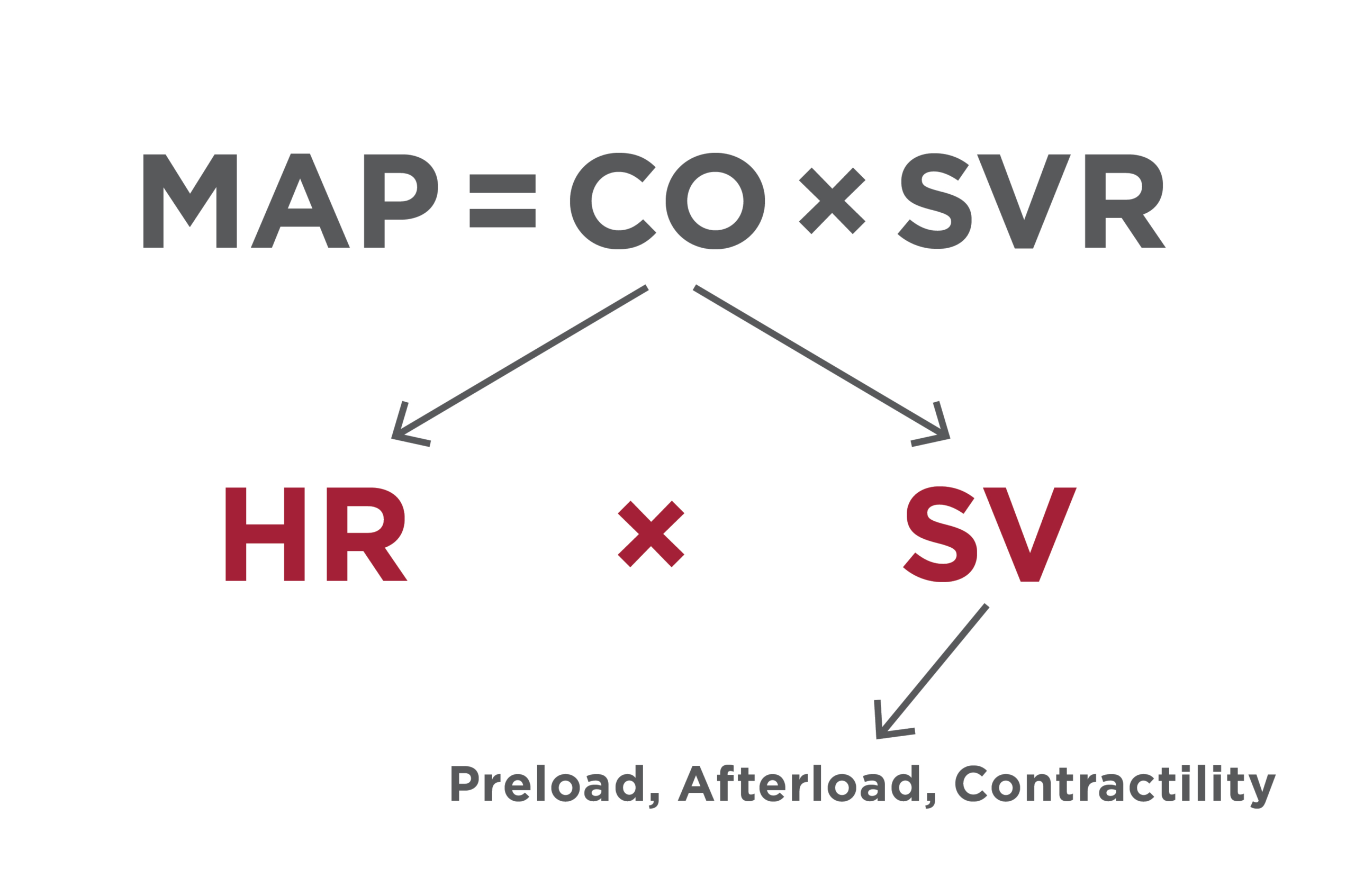
Figure 1. Factors contributing to MAP.
CO = cardiac output; HR = heart rate; MAP = mean arterial pressure; SV = stroke volume; SVR = systemic vascular resistance
Maintaining Normotension
The appropriate therapy to support blood pressure during anesthesia depends on which determinant of MAP is decreased (FIGURE 1).
- Heart rate: Anticholinergics, such as atropine and glycopyrrolate, are useful when bradycardia is contributing to hypotension. They block vagal influence on the sinoatrial and atrioventricular nodes, increasing heart rate.1,4
- Cardiac output: Inotropes and vasopressors may be necessary to support blood pressure. Inotropes like dobutamine improve cardiac contractility and cardiac output. Vasopressors such as norepinephrine and vasopressin increase SVR. Mixed agents like dopamine and ephedrine provide dose-dependent effects on both the heart and vessels by working on α and β receptors. These drugs require careful monitoring, as they can alter organ perfusion, trigger arrhythmias, or lose effectiveness over time.2
- Decreased preload: Fluid therapy is essential when decreased preload or hypovolemia is present. Administering a fluid bolus can restore intravascular volume, improving venous return and stroke volume and ultimately supporting cardiac output and blood pressure. However, if preload is already adequate, excessive fluid administration may lead to fluid overload, tissue edema, and impaired oxygen delivery. For this reason, veterinary professionals providing anesthesia should use caution when administering fluid boluses, particularly in patients with cardiac disease, pulmonary pathology, or other conditions that predispose them to fluid overload.
Hypoventilation
Hypoventilation occurs when the patient cannot eliminate carbon dioxide effectively, resulting in hypercapnia, or a Paco2 (partial pressure of arterial carbon dioxide) above the normal range of 35 to 45 mm Hg. This value is typically obtained via an arterial blood gas measurement.
Carbon dioxide is acidic, and as Paco₂ increases, blood pH decreases because carbon dioxide combines with water to form carbonic acid, leading to respiratory acidosis. In anesthetized patients, end-tidal carbon dioxide (ETco₂) provides a noninvasive estimate of Paco₂ and is typically 2 to 5 mm Hg lower than Paco₂ in patients with normal ventilation and perfusion. The normal ETco₂ range is 35 to 45 mm Hg, corresponding to a normal arterial pH of approximately 7.35 to 7.45. For example, a dog with an ETco₂ of 60 mm Hg would likely have a Paco₂ of approximately 62 to 65 mm Hg and a blood pH around 7.2. This acidosis can depress myocardial contractility, alter electrolyte balance, and increase the risk of cardiac arrhythmias. Severe acidosis can impair cellular function, and significant cell injury may occur when pH falls to approximately 6.8 to 7. As a guideline, for every 10–mm Hg increase in Paco₂ above 40 mm Hg, blood pH decreases by approximately 0.08 to 0.1 pH units during acute respiratory acidosis.6
During recovery, the alveolar gas equation can help quantify the effects of hypoventilation on oxygenation (BOX 1). Alveolar oxygen partial pressure (Pao₂) represents the amount of oxygen available within the alveoli for diffusion into the pulmonary capillary blood. Because Pao₂ is the driving force for oxygen transfer into the bloodstream, decreases in Pao₂ can lead to reduced arterial oxygenation and hypoxemia. Changes in the fraction of inspired oxygen (Fio₂) have a dramatic effect on Pao₂, highlighting how hypoventilation and other physiologic variables can alter oxygen availability and affect oxygenation.3
This equation calculates the partial pressure of alveolar oxygen (Pao2). The fraction of inspired oxygen (Fio2) is 0.21 in room air but depends on the oxygen source. Barometric pressure is 760 mm Hg at sea level but depends on the local elevation. Water vapor pressure in the alveoli is 47 mm Hg at normal body temperature (37 °C [98.6 °F]) and is consistent across human and veterinary species because it is determined by the physical properties of water at body temperature. The respiratory quotient (R) is usually 0.8. Normal Pao2 when breathing room air (21% Fio2) is approximately 100 mm Hg.
Both partial pressure of arterial oxygen (Paco2 ) and Fio2 have significant effects on Pao2. For example, if Paco2 is normal (e.g., 40 mm Hg) in a patient breathing room air at sea level:
Pao2 = 0.21 × (760 − 47 ) − (40 / 0.8)
Pao2 = 0.21 × 713 – 50 = 100 mm Hg
However, if the Pao2 is elevated to 65 mm Hg in the same patient:
Pao2 = 0.21 × (760 − 47) − (65 / 0.8)
Pao2 = 0.21 × 713 – 81 = 69 mm Hg
Elevating the Fio2 in this patient results in:
Pao2 = 0.3 × (760 − 47) − (65 / 0.8)
Pao2 = 0.3 × 713 – 81 = 133 mm Hg
Ventilation Versus Respiratory Rate
When assessing ventilation, it is important to differentiate it from respiratory rate. Respiratory rate refers only to the number of breaths taken per minute, whereas ventilation (minute ventilation) represents the total volume of air moved in and out of the lungs each minute and is calculated as tidal volume (Vt) multiplied by respiratory rate. Because both variables influence ventilation, a patient may have a normal respiratory rate but still be hypoventilating if tidal volume is inadequate.7
Although a low respiratory rate is often assumed to indicate hypoventilation, this is not always the case; ventilation should instead be assessed by evaluating carbon dioxide levels and overall breathing effectiveness. When determining whether a patient’s ventilation is appropriate, it is helpful to consider what the respiratory rate would be if the patient were truly resting or sleeping.2,6
Potential Causes of Hypoventilation Under Anesthesia
- Drug-induced: Anesthetic agents such as opioids, propofol, alfaxalone, and inhalants produce dose-dependent respiratory depression, including apnea or hypoventilation. In a patient without underlying respiratory disease, Paco2 typically remains around 40 mm Hg.2,6 Administration of opioid boluses increases the central nervous system threshold for carbon dioxide, reducing the drive to breathe and promoting hypercapnia.
- Obesity: Patients with a high body condition score are at greater risk for hypoventilation due to excess pressure on the intercostal muscles, impairing full chest expansion. Positioning these patients in sternal recumbency whenever possible allows optimal lung expansion.2,8 When providing mechanical ventilation in obese patients, tidal volume should be calculated as Vt = body weight (kg) × 10 to 20 mL, which often requires higher peak inspiratory pressures on the ventilator.6,9
- Physiologic constraints: Pulmonary disease, muscle fatigue (e.g., myasthenia gravis), or surgical manipulation of the phrenic nerve can limit effective ventilation and predispose patients to hypercapnia.
Maintaining Ventilation
Combating hypoventilation during recovery centers on supporting both ventilation and oxygenation while the patient regains adequate respiratory drive. If the patient becomes hypoxemic or hypercapnic, reintubation with manual or mechanical ventilation may be necessary. When intubation is not required, supplemental oxygen should be readily available to increase the Fio2 and be provided via flow-by, face mask, or oxygen cage, with the patient maintained in sternal recumbency to promote optimal lung expansion. Reversal of respiratory-depressant drugs, such as opioids, can be considered when appropriate but only after confirming the patient is not experiencing pain.
If a patient becomes hypoxemic after extubation but improves with supplemental oxygen (increasing the Fio2), the cause is often residual hypoventilation. In these cases, management should balance ventilatory support with adequate analgesia, ensuring the patient does not require sedation or pain control before administering any reversal agents.
Arrhythmias
Cardiac arrhythmias during anesthesia can range from subtle and clinically insignificant to life-threatening. Understanding the type of arrhythmia and its underlying cause is critical in deciding whether intervention is necessary.
Common Arrhythmias
- Sinus bradycardia: Often drug-induced (e.g., α2 agonists); may not require treatment unless cardiac output is compromised.
- Sinus arrhythmia: A benign variation caused by fluctuations in vagal tone with respiration. It is common and usually not concerning unless the patient is hemodynamically compromised.
- Atrioventricular blocks:
- First degree: Prolonged PR interval; generally hard to diagnose and asymptomatic.
- Second degree: Some P waves fail to trigger a QRS complex; may require monitoring or treatment with an anticholinergic if hemodynamically affected. Dexmedetomidine is known to cause second-degree block; this does not require treatment unless it is affecting the blood pressure negatively.10
- Third degree: Complete dissociation between atrial and ventricular activity; ventricles beat independently, often requiring urgent intervention with anticholinergic or surgical treatment to install a pacemaker.
- Ventricular escape beats versus ventricular premature complexes (VPCs):
- Ventricular escape: A rescue rhythm occurring in response to severe bradycardia. Treatment with an anticholinergic may be required if the patient is hemodynamically compromised.
- VPCs: Often associated with myocardial irritation, hypoxia, or electrolyte disturbances. VPCs may not require treatment unless they are frequent, multifocal, or associated with hemodynamic instability.
Managing Arrhythmias
Effective management of arrhythmias during anesthesia involves identifying the underlying cause, assessing hemodynamic impact, and choosing an appropriate intervention. Continuous monitoring via electrocardiography is essential for early detection and tracking changes in rhythm.
- Assess patient stability: Determine if the arrhythmia is affecting cardiac output or blood pressure. Some arrhythmias are self-limiting and may only require close monitoring.
- Correct underlying factors: Address contributing factors such as hypoxemia, hypercapnia, electrolyte imbalances, acid–base disturbances, or excessive anesthetic depth.
- Pharmacologic intervention: When indicated, anticholinergics such as atropine or glycopyrrolate may be administered to increase heart rate. Antiarrhythmic drugs such as lidocaine may be used for specific ventricular arrhythmias but only after evaluating hemodynamic impact and underlying causes.
- Adjust anesthetic management: Reduce or change drugs that may be contributing to the arrhythmia, such as α2 agonists or inhalant anesthetics. Consider balancing anesthetic depth with analgesia and sedation to minimize cardiovascular depression.
- Supportive care: Maintain normothermia, euhydration, and appropriate patient positioning to reduce stress on the cardiovascular system. For severe or refractory arrhythmias, advanced interventions such as temporary transthoracic pacing, electrical therapy, or referral to a specialty facility may be necessary.
Pain
Patients under general anesthesia do not perceive pain because the anesthetic state suppresses awareness. However, nociception (the neural processing of noxious stimuli) can still occur during general anesthesia. Although these patients may not show overt behavioral signs of pain, inadequate analgesia can still trigger a sympathetic response, resulting in increased heart rate and blood pressure. This stimulation of the nociceptors may originate from surgical manipulation or even patient positioning, particularly in individuals with conditions such as osteoarthritis.
The Pain Pathway
The pain pathway begins with transduction, in which a noxious stimulus (e.g., toe pinch, surgical incision) is converted into an electrical signal at the level of peripheral nociceptors. This signal is then carried through nerve fibers that relay the impulse to the spinal cord (transmission). At the spinal cord, the signal undergoes modulation, where it may be amplified or inhibited by various neurotransmitters and analgesic drugs. The final stage is perception, which occurs in the brain and results in the conscious awareness of pain, often producing a behavioral response such as withdrawal or vocalization (FIGURE 2).
The earlier steps of the pain pathway—transduction, transmission, and modulation—can still occur in animals under anesthesia. If these signals are not adequately controlled, they can lead to central sensitization, also known as windup, in which the nervous system becomes more responsive to pain. This may result in increased postoperative discomfort, delayed recovery, and a higher risk of chronic pain development.11,12
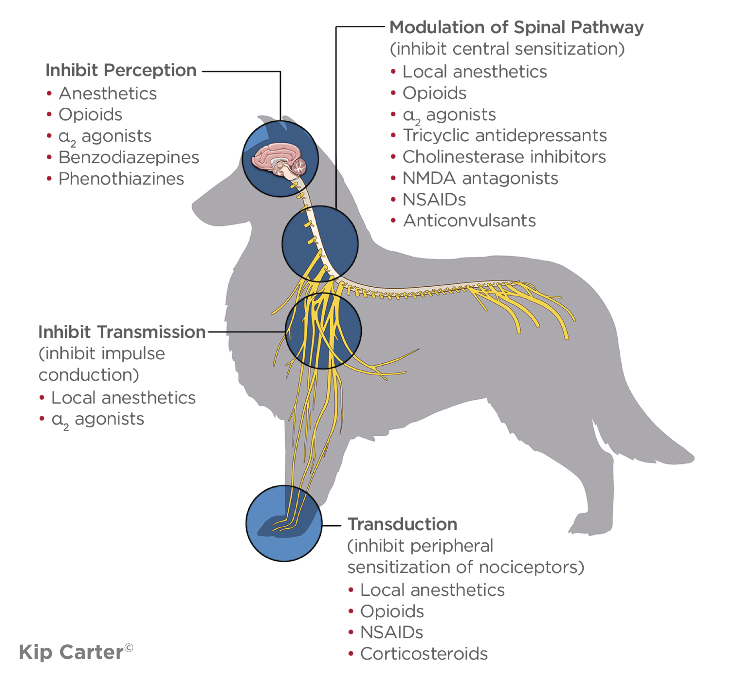
Figure 2. Sites of action of analgesic drugs within the pain pathway. Nociception involves 4 sequential processes: transduction, transmission, modulation, and perception. Analgesic drugs exert their effects by targeting one or more of these stages to reduce pain signaling. Combining medications with different mechanisms of action provides multimodal analgesia, resulting in improved pain control while minimizing the dose and potential adverse effects of individual drugs.
NMDA = N-methyl-D-aspartate
Managing Pain
It is vital to provide all patients with a multimodal analgesic plan that targets different points along the pain pathway. This approach improves patient comfort and reduces reliance on any single drug class.
Common components of multimodal analgesia include:
- NSAIDs to provide peripheral anti-inflammatory and analgesic effects by blunting transmission to the spinal cord
- Local or regional blocks to prevent transmission of nociceptive signals at the surgical site
- Opioids to provide central analgesia by acting on receptors in the brain and spinal cord
- Nonpharmacologic interventions, including patient positioning, gentle handling, cryotherapy, massage, and environmental comfort measures
Studies in veterinary patients have shown that appropriate perioperative analgesia improves comfort, reduces stress responses, and promotes faster recovery.11,13 Effective pain management before, during, and after surgery is essential for reducing patient stress, improving healing, and preventing central sensitization.11,12,14
Hypothermia
Hypothermia is a common complication during anesthesia, especially in small, pediatric, geriatric, or hemodynamically unstable patients. Normal body temperature in dogs and cats generally ranges from 37.5 °C to 39.2 °C (99.5 °F to 102.6 °F). Mild hypothermia occurs below 36.5 °C (97.7 °F), while temperatures below 35 °C (95 °F) can significantly impair cardiovascular and metabolic function.10,15 Decreases in body temperature can have several negative effects, including delayed anesthetic recovery, impaired coagulation, suppressed immune function, and postoperative shivering. Shivering is concerning because it is painful and can increase oxygen consumption by up to 400%, placing additional strain on the cardiovascular and respiratory systems.10
Preventive strategies should focus on maintaining normothermia throughout the perioperative period. This includes using forced-air warming devices, minimizing the use of wetting agents such as alcohol, limiting surgical time when possible, and warming the patient before induction. Careful temperature monitoring and active warming measures are essential for reducing hypothermia-related complications and promoting smoother and faster recoveries.10,15
Thermal injury can occur when tissues are exposed to temperatures above approximately 43 °C to 45 °C (109 °F to 113 °F) for sustained periods. Anesthetized patients are particularly vulnerable because they are unable to perceive heat or move away from the source. Direct-contact warming methods, such as microwaved heat packs, electric heating pads, or hot water bottles have been associated with thermal burns in veterinary patients. For this reason, active warming systems designed for anesthesia, such as forced-air warming units or circulating warm-water blankets, are recommended as safer and more controlled options.10
Role of the Veterinary Nurse
Veterinary nurses are often the primary anesthesia providers in veterinary practice, making their knowledge and decision-making skills critical to patient safety. Well-trained veterinary nurses understand how anesthetic drugs affect each body system, how to interpret monitoring parameters, and how to intervene appropriately when abnormalities arise. This depth of knowledge transforms monitoring from simply watching numbers on a screen into active, informed patient care. Ongoing education and direct communication with the veterinarian overseeing the anesthesia, hands-on training, and familiarity with equipment and protocols empower veterinary nurses to respond confidently and effectively in challenging situations. From adjusting anesthetic depth to initiating ventilatory support or addressing hypotension, the veterinary nurse’s actions directly influence anesthetic stability and recovery quality.
Summary
Understanding and managing common anesthetic complications such as hypotension, hypoventilation, arrhythmias, pain, and hypothermia are essential for effective patient safety during anesthesia. Each of these conditions requires careful assessment and timely intervention. Knowledge truly is power in anesthesia; the more prepared and educated the veterinary nurse, the safer the anesthetic experience will be for the patient.
References
- Penev Y, Ruppert MM, Bilgili A, et al. Intraoperative hypotension and postoperative acute kidney injury: a systematic review. Am J Surg. 2024;232:45-53. doi:10.1016/j.amjsurg.2024.02.001
- Muir WW. Cardiovascular physiology and pathophysiology. In: Grimm KA, Lamont LA, Tranquilli WJ, Greene SA, Robertson SA, eds. Lumb and Jones’ Veterinary Anesthesia and Analgesia. 6th ed. Wiley-Blackwell; 2024:615-666.
- Hall JE, Hall ME. Cardiac output, venous return, and their regulation. In: Hall JE, Hall ME, eds. Guyton and Hall Textbook of Medical Physiology. 14th ed. Elsevier; 2021:245-258.
- Bruniges N, Rioja E. Intraoperative anaesthetic complications in dogs undergoing general anaesthesia for thoracolumbar hemilaminectomy: a retrospective analysis. Vet Anaesth Analg. 2019;46(6):720-728.
- Hall JE, Hall ME. Arterial pressure and hypertension. In: Hall JE, Hall ME, eds. Guyton and Hall Textbook of Medical Physiology. 14th ed. Elsevier; 2021:489-540.
- Muir WW. Respiratory physiology and pulmonary function. In: Grimm KA, Lamont LA, Tranquilli WJ, Greene SA, Robertson SA, eds. Lumb and Jones’ Veterinary Anesthesia and Analgesia. 6th ed. Wiley-Blackwell; 2024:697-749.
- Hall JE, Hall ME. Respiratory physiology. In: Hall JE, Hall ME, eds. Guyton and Hall Textbook of Medical Physiology. 14th ed. Elsevier; 2021:511-520.
- Hofmeister EH. Anesthetic emergencies, resuscitation, and adverse events. In: Grimm KA, Lamont LA, Tranquilli WJ, Greene SA, Robertson SA, eds. Lumb and Jones’ Veterinary Anesthesia and Analgesia. 6th ed. Wiley-Blackwell; 2024:54-73.
- Dugdale A. Respiratory considerations. In: Dugdale A, Beaumont G, Bradbrook C, Gurney M, eds. Veterinary Anaesthesia: Principles to Practice. 2nd ed. Wiley-Blackwell; 2020:607-610.
- Creighton CM, Johnson Bressan N. Monitoring the anesthetized patient. In: Grimm KA, Lamont LA, Tranquilli WJ, Greene SA, Robertson SA. Lumb and Jones’ Veterinary Anesthesia and Analgesia. 6th ed. Wiley-Blackwell; 2024:169-175.
- McKune CM. Clinical management and pharmacologic treatment of pain. In: Grimm KA, Lamont LA, Tranquilli WJ, Greene SA, Robertson SA, eds. Lumb and Jones’ Veterinary Anesthesia and Analgesia. 6th ed. Wiley-Blackwell; 2024:1010-1022.
- Mathews KA, Kronen PW, Lascelles D, et al. Guidelines for recognition, assessment and treatment of pain. J Small Anim Pract. 2014;55(6):E10-E68. https://doi.org/10.1111/jsap.12200
- Gaynor JS, Muir WW. Physiology and pathophysiology of pain. In: Gaynor JS, Muir WW, eds. Handbook of Veterinary Pain Management. 2nd ed. Mosby/Elsevier; 2008:13-41.
- Gaynor JS, Muir WW. Objective, categoric methods for assessing pain and analgesia. In: Gaynor JS, Muir WW, eds. Handbook of Veterinary Pain Management. 2nd ed. Mosby/Elsevier; 2008:78-109.
- Sessler DI. Complications and treatment of mild hypothermia. Anesthesiology. 2001:95(2):531-543. doi:10.1097/00000542-200108000-00040
CE Quiz
This article has been submitted for RACE approval for 1 hour of continuing education credit and will be opened for enrollment upon approval. To receive credit, take the test at vetfolio.com. Free registration is required. Questions and answers online may differ from those below. Tests are valid for 2 years from the date of approval.
1. What mean arterial pressure (MAP) is generally considered the minimum acceptable value to maintain adequate organ perfusion in anesthetized patients?
a. 40 mm Hg
b. 50 mm Hg
c. 60–70 mm Hg
d. 80–90 mm Hg
2. An end-tidal carbon dioxide (ETco2) reading of 65 mm Hg during anesthesia most likely indicates:
a. Hyperventilation
b. Hypoventilation
c. Normal ventilation
d. Equipment malfunction
3. Which of the following is a common consequence of perioperative hypothermia?
a. Faster anesthetic recovery
b. Increased immune response
c. Delayed recovery and impaired coagulation
d. Decreased oxygen consumption
4. Why is multimodal analgesia recommended in anesthetized patients?
a. It reduces the need for monitoring.
b. It targets different points in the pain pathway.
c. It eliminates the need for opioids.
d. It prevents hypothermia.
5. MAP depends on which 2 factors?
a. Cardiac output and systemic vascular resistance
b. Blood pressure and heart rate
c. Pain and anesthetic depth
d. Hypothermia and pain